Abstract
BACKGROUND/AIMS—Periventricular leucomalacia (PVL) is a lesion in the immature brain involving the optic radiation. Children with PVL have visual problems including crowding, visual field defects, strabismus, and visual perceptual/cognitive deficits, together with nystagmus. They often have optic nerve hypoplasia seen either as small discs or as large cupping of normal sized optic discs. This study aimed to perform eye movement recordings in a group of children with PVL in order to characterise and classify the nystagmus. METHODS—19 children with PVL on cerebral imaging underwent eye movement recordings with the Ober-2 infrared reflection technique. RESULTS—16 of the 19 subjects had horizontal nystagmus. CONCLUSION—The present study shows that nystagmus is commonly seen in children with PVL. Keywords: nystagmus; periventricular leucomalacia; visual impairment; cerebral imaging; eye movement; infrared reflection technique
Full Text
The Full Text of this article is available as a PDF (141.0 KB).
Figure 1 .
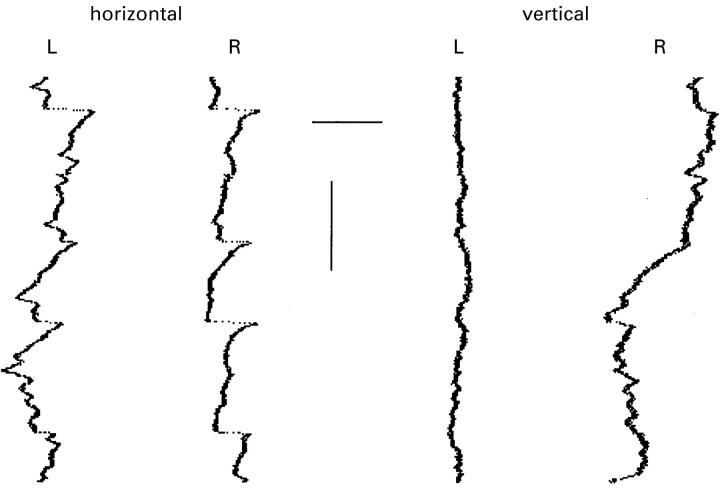
Eye movement record showing horizontal and vertical traces of the right (R) and left (L) eyes with the Ober-2 system. Patient no 9 when fixating in primary position with both eyes open. Note the variable amplitude and the asymmetrical waveforms with a SPD (slow phase decreasing velocity) waveform most visible in the right eye trace. Note also the difference in vertical stability between the right and left eye. Horizontal bar, 3 degrees; vertical bar, 500 ms.
Figure 2 .
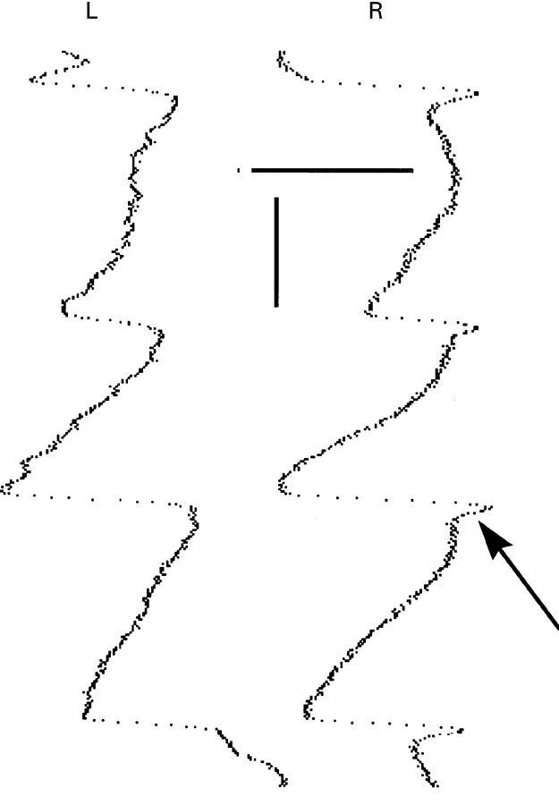
Eye movement record showing horizontal and vertical traces of the right (R) and left (L) eyes with the Ober-2 system. Patient no 2 with only horizontal traces shown. The record showing a jerk nystagmus with a SPI (slow phase exponentially increasing velocity) waveform with a superimposed foveating saccade (arrow). Horizontal bar, 2 degrees; vertical bar, 200 ms.
Figure 3 .
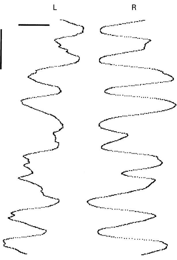
Eye movement record showing horizontal and vertical traces of the right (R) and left (L) eyes with the Ober-2 system. Patient no 12 with a asymmetrical pendular nystagmus out of phase. Horizontal bar, 4 degrees; vertical bar, 500 ms.
Figure 4 .

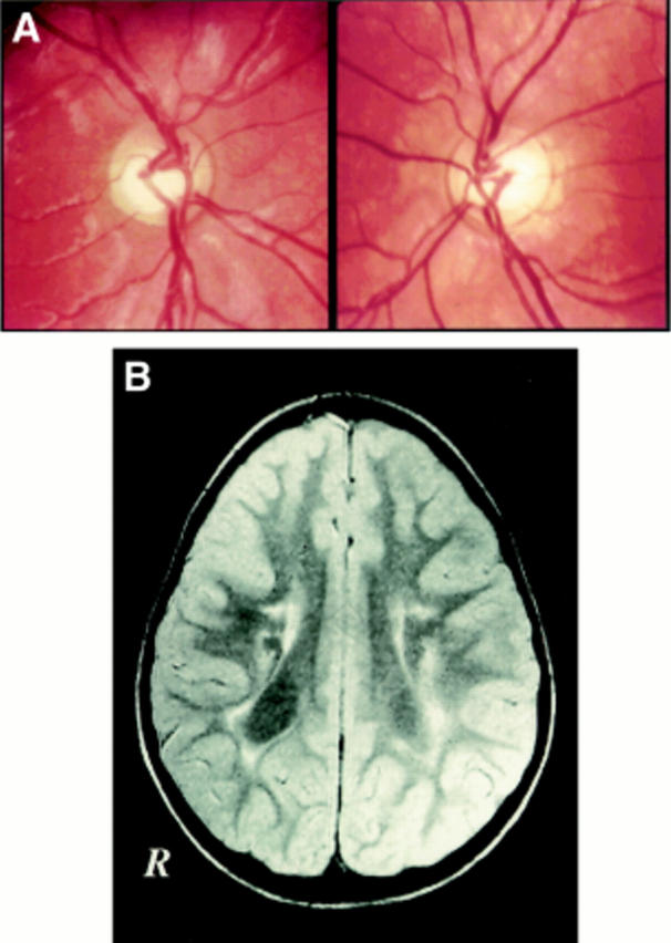
(A) Fundus photograph of patient no 9 demonstrating large cups in normal sized discs. (B) This single MRI shows typical changes of periventricular leucomalacia with atrophic dilatation of the posterior parts of the lateral ventricles. The amount of periventricular white matter is reduced around the occipital horns and adjacent to the trigone bilaterally. Remaining white matter has an abnormally bright signal on this T2 weighted image, first echo, indicating permanent damage, gliosis. Variations of signal involving left posterior cortical structures represent artefacts in the image.
Selected References
These references are in PubMed. This may not be the complete list of references from this article.
- Abadi R. V., Dickinson C. M. Waveform characteristics in congenital nystagmus. Doc Ophthalmol. 1986;64(2):153–167. doi: 10.1007/BF00159990. [DOI] [PubMed] [Google Scholar]
- BANKER B. Q., LARROCHE J. C. Periventricular leukomalacia of infancy. A form of neonatal anoxic encephalopathy. Arch Neurol. 1962 Nov;7:386–410. doi: 10.1001/archneur.1962.04210050022004. [DOI] [PubMed] [Google Scholar]
- Dell'Osso L. F. Congenital, latent and manifest latent nystagmus--similarities, differences and relation to strabismus. Jpn J Ophthalmol. 1985;29(4):351–368. [PubMed] [Google Scholar]
- Dell'Osso L. F., Daroff R. B. Congenital nystagmus waveforms and foveation strategy. Doc Ophthalmol. 1975 Nov 21;39(1):155–182. doi: 10.1007/BF00578761. [DOI] [PubMed] [Google Scholar]
- Dutton G., Ballantyne J., Boyd G., Bradnam M., Day R., McCulloch D., Mackie R., Phillips S., Saunders K. Cortical visual dysfunction in children: a clinical study. Eye (Lond) 1996;10(Pt 3):302–309. doi: 10.1038/eye.1996.64. [DOI] [PubMed] [Google Scholar]
- Eken P., van Nieuwenhuizen O., van der Graaf Y., Schalij-Delfos N. E., de Vries L. S. Relation between neonatal cranial ultrasound abnormalities and cerebral visual impairment in infancy. Dev Med Child Neurol. 1994 Jan;36(1):3–15. doi: 10.1111/j.1469-8749.1994.tb11760.x. [DOI] [PubMed] [Google Scholar]
- Farmer J., Hoyt C. S. Monocular nystagmus in infancy and early childhood. Am J Ophthalmol. 1984 Oct 15;98(4):504–509. doi: 10.1016/0002-9394(84)90140-5. [DOI] [PubMed] [Google Scholar]
- Gelbart S. S., Hoyt C. S. Congenital nystagmus: a clinical perspective in infancy. Graefes Arch Clin Exp Ophthalmol. 1988;226(2):178–180. doi: 10.1007/BF02173313. [DOI] [PubMed] [Google Scholar]
- Good W. V., Jan J. E., DeSa L., Barkovich A. J., Groenveld M., Hoyt C. S. Cortical visual impairment in children. Surv Ophthalmol. 1994 Jan-Feb;38(4):351–364. doi: 10.1016/0039-6257(94)90073-6. [DOI] [PubMed] [Google Scholar]
- Gresty M. A., Metcalfe T., Timms C., Elston J., Lee J., Liu C. Neurology of latent nystagmus. Brain. 1992 Oct;115(Pt 5):1303–1321. doi: 10.1093/brain/115.5.1303. [DOI] [PubMed] [Google Scholar]
- Hagberg B., Hagberg G., Olow I. The changing panorama of cerebral palsy in Sweden 1954-1970. I. Analysis of the general changes. Acta Paediatr Scand. 1975 Mar;64(2):187–192. doi: 10.1111/j.1651-2227.1975.tb03820.x. [DOI] [PubMed] [Google Scholar]
- Jacobson L., Ek U., Fernell E., Flodmark O., Broberger U. Visual impairment in preterm children with periventricular leukomalacia--visual, cognitive and neuropaediatric characteristics related to cerebral imaging. Dev Med Child Neurol. 1996 Aug;38(8):724–735. doi: 10.1111/j.1469-8749.1996.tb12142.x. [DOI] [PubMed] [Google Scholar]
- Jacobson L., Hellström A., Flodmark O. Large cups in normal-sized optic discs: a variant of optic nerve hypoplasia in children with periventricular leukomalacia. Arch Ophthalmol. 1997 Oct;115(10):1263–1269. doi: 10.1001/archopht.1997.01100160433007. [DOI] [PubMed] [Google Scholar]
- Krägeloh-Mann I., Petersen D., Hagberg G., Vollmer B., Hagberg B., Michaelis R. Bilateral spastic cerebral palsy--MRI pathology and origin. Analysis from a representative series of 56 cases. Dev Med Child Neurol. 1995 May;37(5):379–397. doi: 10.1111/j.1469-8749.1995.tb12022.x. [DOI] [PubMed] [Google Scholar]
- Schenk-Rootlieb A. J., Van Nieuwenhuizen O., Schiemanck N., Van der Graaf Y., Willemse J. Impact of cerebral visual impairment on the everyday life of cerebral palsied children. Child Care Health Dev. 1993 Nov-Dec;19(6):411–423. doi: 10.1111/j.1365-2214.1993.tb00745.x. [DOI] [PubMed] [Google Scholar]
- Schenk-Rootlieb A. J., van Nieuwenhuizen O., van der Graaf Y., Wittebol-Post D., Willemse J. The prevalence of cerebral visual disturbance in children with cerebral palsy. Dev Med Child Neurol. 1992 Jun;34(6):473–480. doi: 10.1111/j.1469-8749.1992.tb11467.x. [DOI] [PubMed] [Google Scholar]
- Tusa R. J., Ungerleider L. G. Fiber pathways of cortical areas mediating smooth pursuit eye movements in monkeys. Ann Neurol. 1988 Feb;23(2):174–183. doi: 10.1002/ana.410230211. [DOI] [PubMed] [Google Scholar]
- Weiss A. H., Biersdorf W. R. Visual sensory disorders in congenital nystagmus. Ophthalmology. 1989 Apr;96(4):517–523. doi: 10.1016/s0161-6420(89)32864-8. [DOI] [PubMed] [Google Scholar]
- Wiklund L. M., Uvebrant P., Flodmark O. Computed tomography as an adjunct in etiological analysis of hemiplegic cerebral palsy. I: Children born preterm. Neuropediatrics. 1991 Feb;22(1):50–56. doi: 10.1055/s-2008-1071417. [DOI] [PubMed] [Google Scholar]


