Abstract
AIMS—To devise a method to measure tear meniscus curvature by a non-invasive specular technique. METHODS—A photographic system was devised. The system consisted of a camera and an illuminated target with a series of black and white stripes oriented parallel to the axis of the lower tear meniscus. The target was mounted on a flash gun close to the objective of a Brown macrocamera and calibrated using a graduated series of glass capillaries of known diameter, ground down to expose the inner wall. It was then applied to normal human eyes (n=45) to measure the tear meniscus curvature. A video system was also assessed which provided qualitative online information about the tear meniscus. RESULTS—Using the photographic system, measured values for capillary radii were in excellent agreement with theoretical calculations (r2=0.996, p<0.0001). The radii of curvature of lower tear menisci in normal human subjects (mean 0.365 (SD 0.153) mm, range 0.128-0.736; n=45) were similar to those reported in the literature. Both systems demonstrated variations in meniscus shape. The video system provided stable images of human menisci over prolonged periods of time and promises to be useful for the analysis of dynamic changes in meniscus volume. CONCLUSIONS—Reflective meniscometry is a non-invasive technique providing quantitative information about tear meniscus shape and volume and of potential value in the study of ocular surface disease. Keywords: tear meniscus; reflective meniscometry; radius of curvature; tear kinetics
Full Text
The Full Text of this article is available as a PDF (147.8 KB).
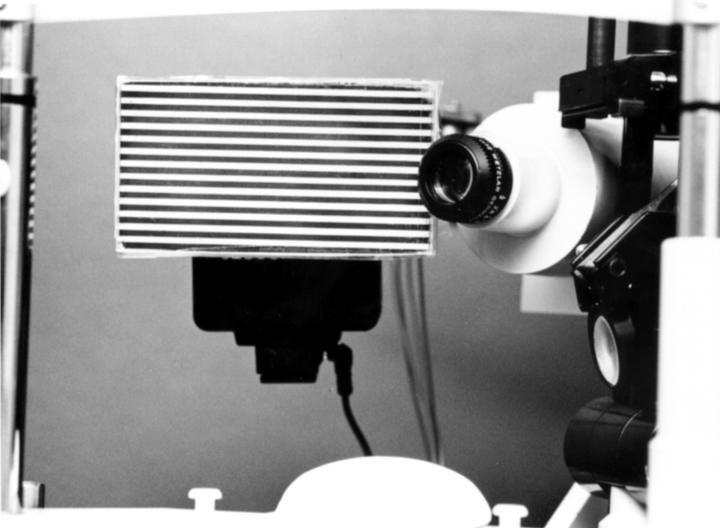
Figure 1 .
Front view of the photographic system, consisting of the target and a Brown macrocamera.
Figure 2 .
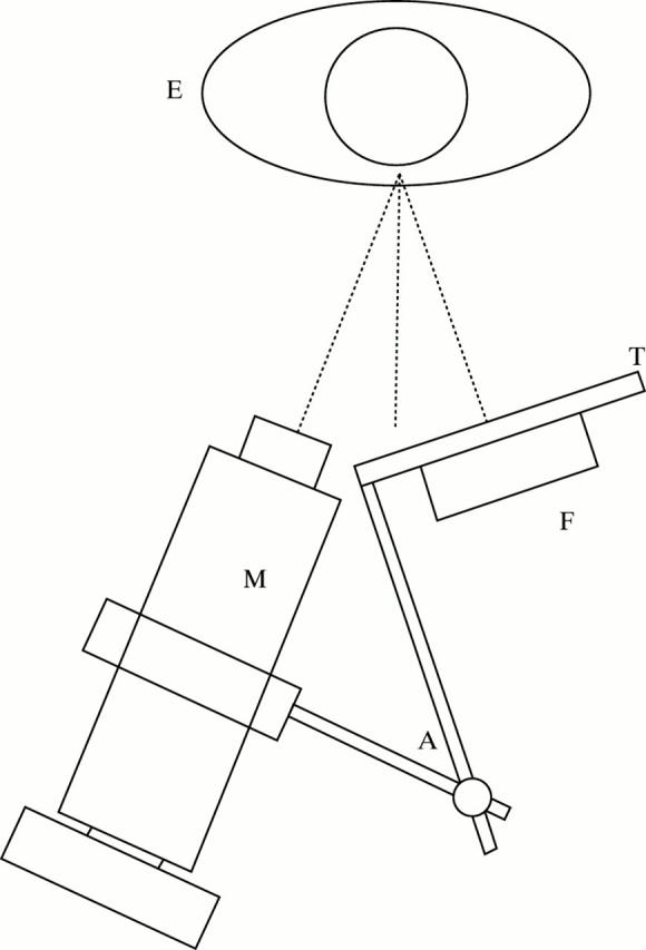
Diagram of the photographic system. E=subject's eye; M=Brown's macrocamera; T=target; F=flash; A=flexible arm.
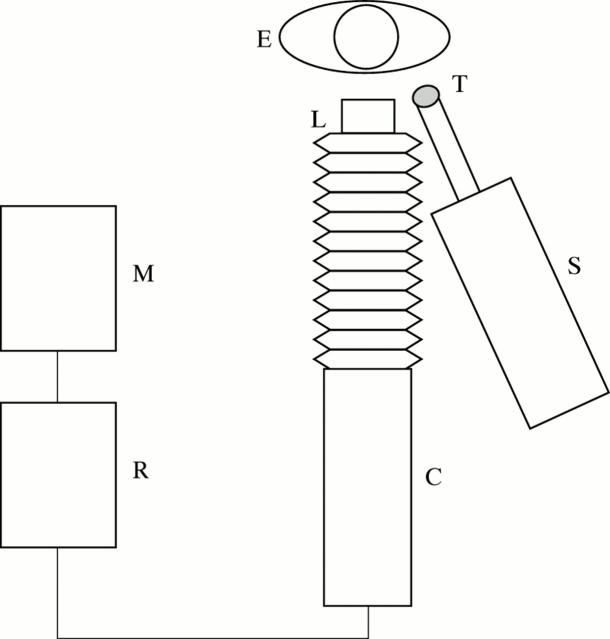
Figure 3 .
Diagram of the video system. E=subject's eye; L=lens; T=target; S=light source; C=video camera; R=video recorder; M=monitor.
Figure 4 .
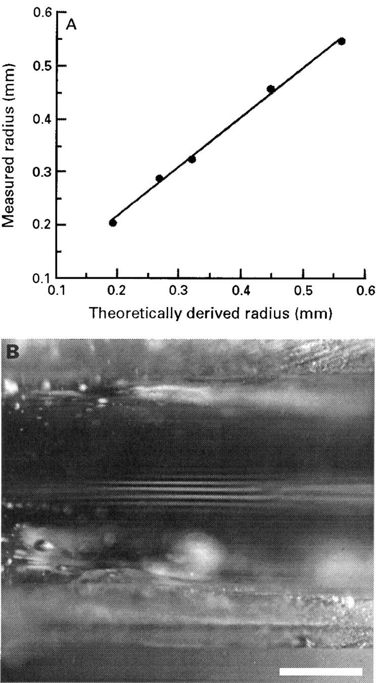
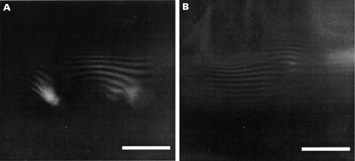
(A) Relation between measured and theoretically derived radius of curvature of an individual capillary tube target. Excellent agreement is shown: (y=0.0300+0.936x (r2=0.996, p<0.0001); where x = theoretical (mm), y = measured (mm)). (B) Image formed by a ground down glass capillary tube (radius 0.547 mm; bar=500 µm).
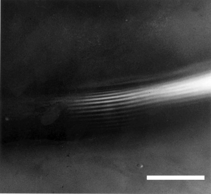
Figure 5 .
A representative example of the normal meniscus of a human eye. Measurement is made in the region of the central part of the lid where the images of the target lines are usually parallel (see text; bar=500 µm).
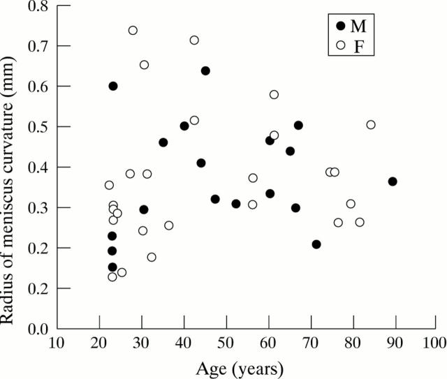
Figure 6 .
Scatter plot of measured radius of curvature against age of subject.
Figure 7 .
Representative examples of variations in the meniscus shape; (A) forward serrations, (B) doubling; bars=500 µm.
Figure 8 .
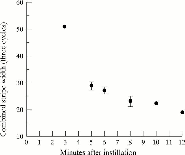
Example showing the change in the tear meniscus curvature, after instillation of a single drop of Celluvisc (1% carboxymethylcellulose sodium, Allergan). Ordinate: Stripe width measured directly from the TV monitor using a multistripe target. The width of three black and white cycles is shown. Abscissa: Time in minutes. Average width (mm) (SD) (error bars are hidden by the symbols at 3, 10, and 12 minutes).
Figure 9 .
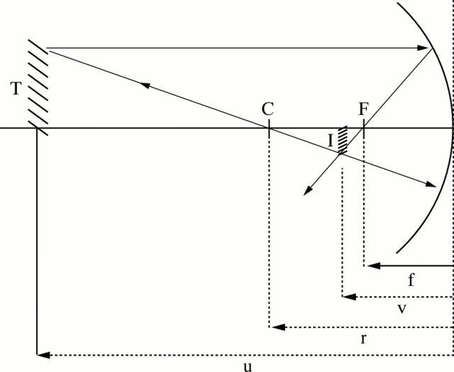
Ray diagram for theoretical determination of the radius of meniscus curvature. T=target; C=centre of the expected circular portion of the meniscus; I=image; F=focal point of the convex meniscus mirror; u=target distance; v=image distance; r=radius of meniscus curvature; f=focal distance.
Selected References
These references are in PubMed. This may not be the complete list of references from this article.
- Brown N. Macrophotography of the anterior segment of the eye. Br J Ophthalmol. 1970 Oct;54(10):697–701. doi: 10.1136/bjo.54.10.697. [DOI] [PMC free article] [PubMed] [Google Scholar]
- Cho P. The cotton thread test: a brief review and a clinical study of its reliability on Hong Kong-Chinese. Optom Vis Sci. 1993 Oct;70(10):804–808. doi: 10.1097/00006324-199310000-00004. [DOI] [PubMed] [Google Scholar]
- Farris R. L., Stuchell R. N., Mandel I. D. Basal and reflex human tear analysis. I. Physical measurements: osmolarity, basal volumes, and reflex flow rate. Ophthalmology. 1981 Aug;88(8):852–857. doi: 10.1016/s0161-6420(81)34939-2. [DOI] [PubMed] [Google Scholar]
- Goren M. B., Goren S. B. Diagnostic tests in patients with symptoms of keratoconjunctivitis sicca. Am J Ophthalmol. 1988 Nov 15;106(5):570–574. doi: 10.1016/0002-9394(88)90587-9. [DOI] [PubMed] [Google Scholar]
- Hamano H., Hori M., Hamano T., Mitsunaga S., Maeshima J., Kojima S., Kawabe H., Hamano T. A new method for measuring tears. CLAO J. 1983 Jul-Sep;9(3):281–289. [PubMed] [Google Scholar]
- Holly F. J., Lemp M. A. Tear physiology and dry eyes. Surv Ophthalmol. 1977 Sep-Oct;22(2):69–87. doi: 10.1016/0039-6257(77)90087-x. [DOI] [PubMed] [Google Scholar]
- Hykin P. G., Bron A. J. Age-related morphological changes in lid margin and meibomian gland anatomy. Cornea. 1992 Jul;11(4):334–342. doi: 10.1097/00003226-199207000-00012. [DOI] [PubMed] [Google Scholar]
- Khurana A. K., Chaudhary R., Ahluwalia B. K., Gupta S. Tear film profile in dry eye. Acta Ophthalmol (Copenh) 1991 Feb;69(1):79–86. doi: 10.1111/j.1755-3768.1991.tb01997.x. [DOI] [PubMed] [Google Scholar]
- Lamberts D. W., Foster C. S., Perry H. D. Schirmer test after topical anesthesia and the tear meniscus height in normal eyes. Arch Ophthalmol. 1979 Jun;97(6):1082–1085. doi: 10.1001/archopht.1979.01020010536004. [DOI] [PubMed] [Google Scholar]
- Loran D. F., French C. N., Lam S. Y., Papas E. Reliability of the wetting value of tears. Ophthalmic Physiol Opt. 1987;7(1):53–56. [PubMed] [Google Scholar]
- Mainstone J. C., Bruce A. S., Golding T. R. Tear meniscus measurement in the diagnosis of dry eye. Curr Eye Res. 1996 Jun;15(6):653–661. doi: 10.3109/02713689609008906. [DOI] [PubMed] [Google Scholar]
- Mathers W. D., Lane J. A., Zimmerman M. B. Tear film changes associated with normal aging. Cornea. 1996 May;15(3):229–234. doi: 10.1097/00003226-199605000-00001. [DOI] [PubMed] [Google Scholar]
- McDonald J. E., Brubaker S. Meniscus-induced thinning of tear films. Am J Ophthalmol. 1971 Jul 30;72(1):139–146. doi: 10.1016/0002-9394(71)91604-7. [DOI] [PubMed] [Google Scholar]
- Mishima S., Gasset A., Klyce S. D., Jr, Baum J. L. Determination of tear volume and tear flow. Invest Ophthalmol. 1966 Jun;5(3):264–276. [PubMed] [Google Scholar]
- Scherz W., Doane M. G., Dohlman C. H. Tear volume in normal eyes and keratoconjunctivitis sicca. Albrecht Von Graefes Arch Klin Exp Ophthalmol. 1974;192(2):141–150. doi: 10.1007/BF00410700. [DOI] [PubMed] [Google Scholar]
- Wong H, Fatt I, I, Radke CJ. Deposition and Thinning of the Human Tear Film. J Colloid Interface Sci. 1996 Dec 1;184(1):44–51. doi: 10.1006/jcis.1996.0595. [DOI] [PubMed] [Google Scholar]
- Zappia R. J., Milder B. Lacrimal drainage function. 1. The Jones fluorescein test. Am J Ophthalmol. 1972 Jul;74(1):154–159. doi: 10.1016/0002-9394(72)91141-5. [DOI] [PubMed] [Google Scholar]
- van Bijsterveld O. P. Diagnostic tests in the Sicca syndrome. Arch Ophthalmol. 1969 Jul;82(1):10–14. doi: 10.1001/archopht.1969.00990020012003. [DOI] [PubMed] [Google Scholar]


