

Full Text
The Full Text of this article is available as a PDF (381.1 KB).
Figure 1 .
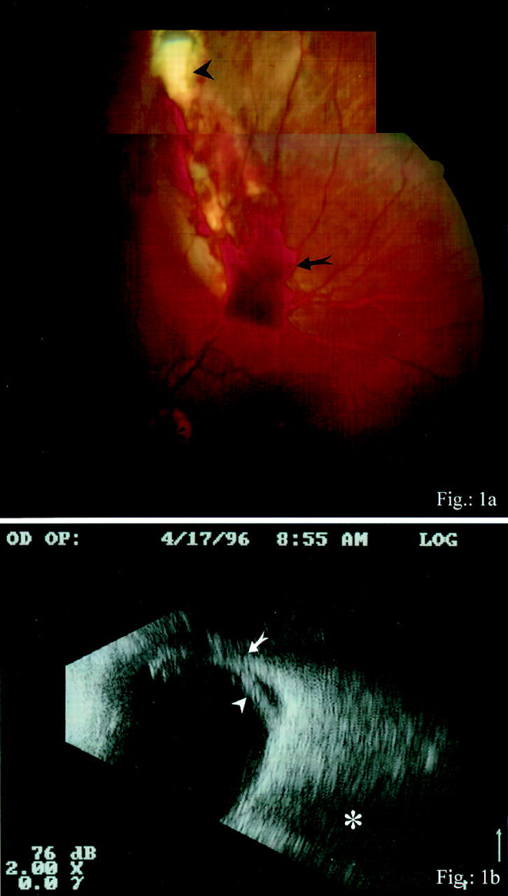
(A) Arrowhead indicates orbital fat in the scleral wound gap. The vitreous haemorrhage is marked with the arrow and the optic nerve head with the asterisk. (B) The arrowhead indicates the detached lamina interna of the vitreous and the arrow shows the area of vitreous incarceration into the scleral wound. The optic nerve is marked with the asterisk.
Figure 2 .
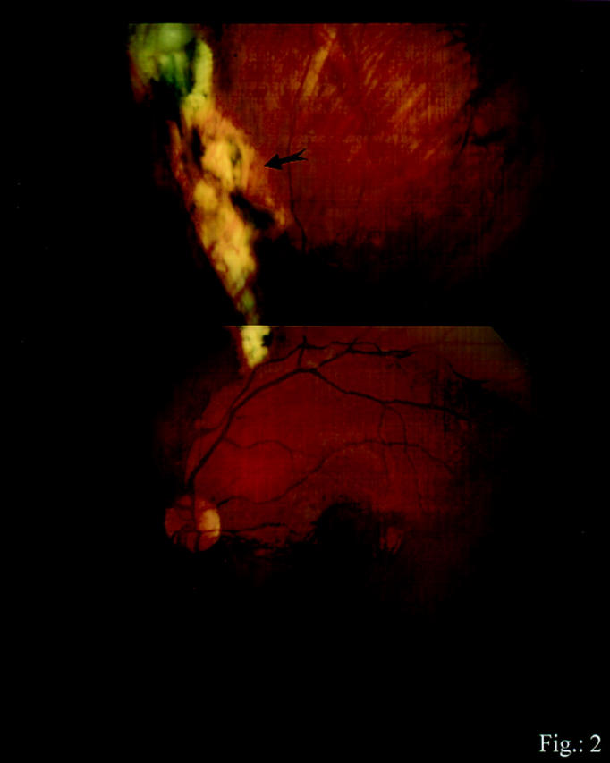
The arrow indicates the huge sclerochorioretinal scar.