Abstract
AIMS—To assess the reproducibility and validity of a new instrument for grading nuclear cataract—the laser slit lamp, by comparison with an established method of lens grading—the Lens Opacities Classification System III (LOCS III). METHODS—62 volunteers (113 eyes) were examined on two occasions. At each visit, a video image of the anterior segment was captured with the laser slit lamp and stored digitally. A measure of lens opacity for each laser slit lamp image was calculated using image analysis software. Each lens was also photographed on both visits for LOCS III grading of nuclear colour and nuclear opalescence. RESULTS—There was a linear increasing relation between the laser slit lamp measure of nuclear lens opacity and the LOCS III nuclear opalescence scores. The overall reproducibility of the laser slit lamp measurement was comparable with LOCS III (intraclass correlation coefficient of 95% (95% CI 92.9-96.5) for the laser slit lamp method; 97% (95% CI 95.6-97.9) for the LOCS III method). However, among healthy subjects, the reproducibility was lower (intraclass correlation for the laser slit lamp of 38.6% (95% CI 12.9-59.2) and 76.1% (95% CI 62.3-85.4) for LOCS III. CONCLUSION—The laser slit lamp appears to give a valid measurement of nuclear cataract. The reproducibility of the instrument was high and similar to that of LOCS III. Modifications to the design would have to be made to improve its reproducibility among healthy subjects. It is simpler than other objective instruments, and could be useful in large scale studies of cataract.
Full Text
The Full Text of this article is available as a PDF (125.2 KB).
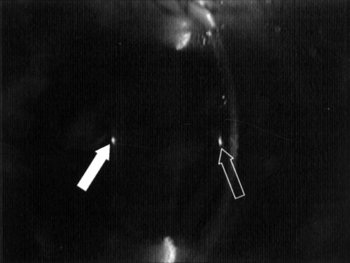
Figure 1 .
Digitised video image of the anterior segment of a normal subject taken with the laser slit lamp. The bright spots are Purkinje images I (open arrow) and IV (solid arrow) and were used to standardise alignment of the eye during image capture.
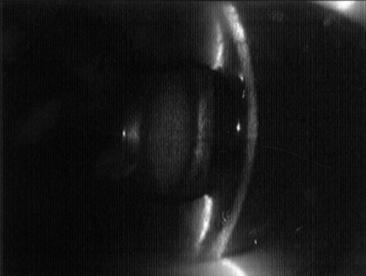
Figure 2 .
Laser slit lamp (digitised video image) view of the anterior segment of a subject with nuclear lens opacity.
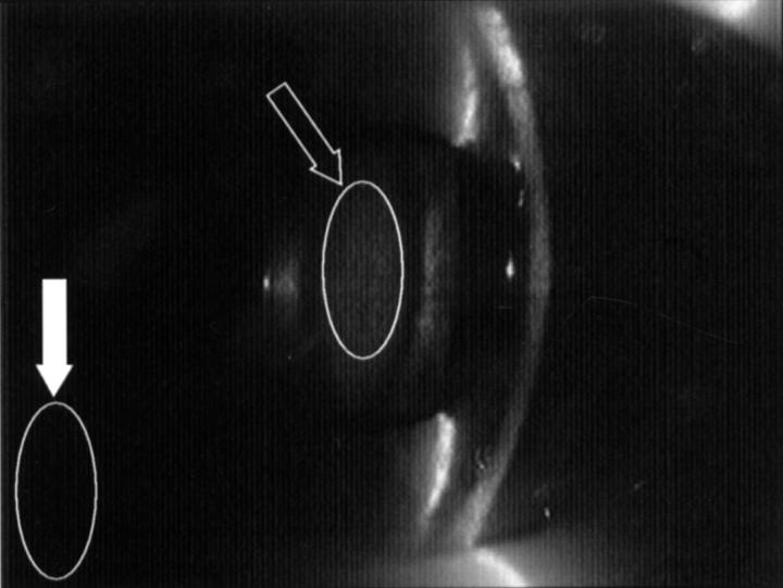
Figure 3 .
Laser slit lamp image showing how the region of interest mask (ellipse) was defined for the nuclear area (open arrow) and for the background area (solid arrow).
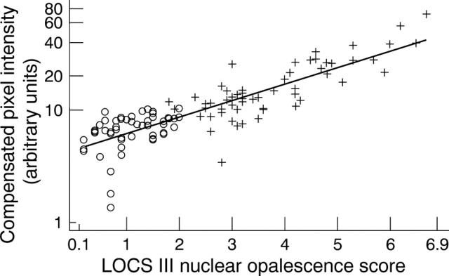
Figure 4 .
Relation between compensated pixel intensities (laser slit lamp) and LOCS III opalescence scores in 55 eyes of healthy subjects (∘), and 58 eyes of subjects from the cataract waiting list (+).
Figure 5 .
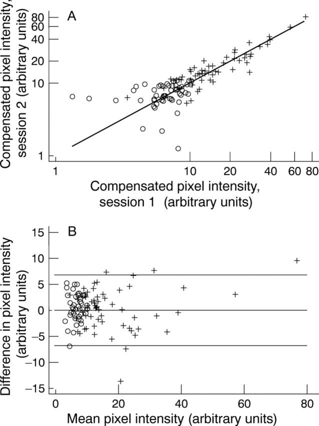
(A) Relation between compensated pixel intensities (laser slit lamp) measured at the first and second visits for 55 eyes of healthy subjects (∘) and 58 eyes of subjects from the cataract waiting list (+). (B) Bland-Altman plot for the laser slit lamp. Difference between the two repeat compensated pixel intensities plotted against the mean of the two readings for 55 eyes of healthy subjects (∘) and 58 eyes of subjects from the cataract waiting list (+).
Figure 6 .
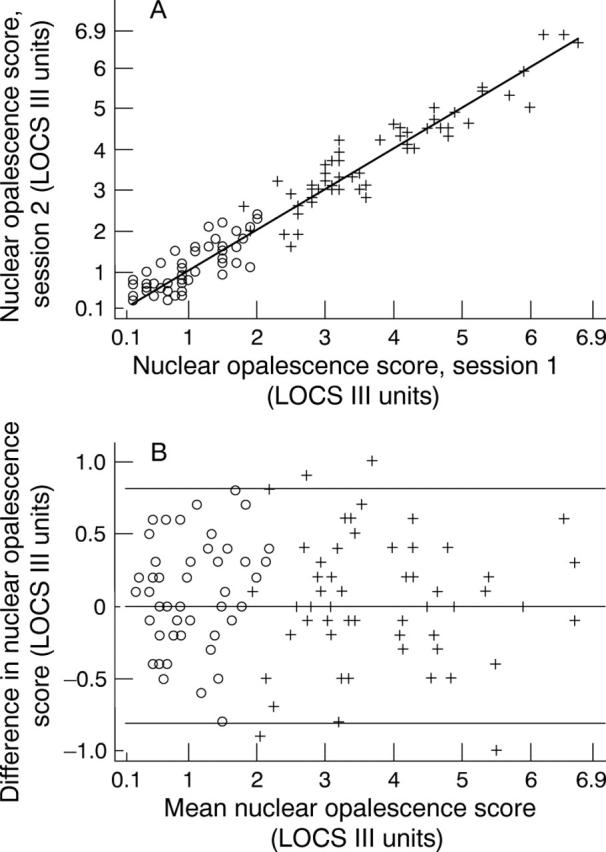
(A) Relation between LOCS III nuclear opalescence scores measured at the first and second visits for 55 eyes of healthy subjects (∘) and 58 eyes of subjects from the cataract waiting list (+). (B) Bland-Altman plot for LOCS III nuclear opalescence. Difference between LOCS III nuclear opalescence scores for the two visits plotted against the mean for 55 eyes of healthy subjects (∘) and 58 eyes of subjects from the cataract waiting list (+).
Selected References
These references are in PubMed. This may not be the complete list of references from this article.
- Brown N. A., Bron A. J., Ayliffe W., Sparrow J., Hill A. R. The objective assessment of cataract. Eye (Lond) 1987;1(Pt 2):234–246. doi: 10.1038/eye.1987.43. [DOI] [PubMed] [Google Scholar]
- Brown N. A., Bron A. J., Sparrow J. M. Methods for evaluation of lens changes. Int Ophthalmol. 1988;12(4):229–235. doi: 10.1007/BF00133938. [DOI] [PubMed] [Google Scholar]
- Chinn S. Statistics in respiratory medicine. 2. Repeatability and method comparison. Thorax. 1991 Jun;46(6):454–456. doi: 10.1136/thx.46.6.454. [DOI] [PMC free article] [PubMed] [Google Scholar]
- Chinn S. The assessment of methods of measurement. Stat Med. 1990 Apr;9(4):351–362. doi: 10.1002/sim.4780090402. [DOI] [PubMed] [Google Scholar]
- Christen W. G., Manson J. E., Seddon J. M., Glynn R. J., Buring J. E., Rosner B., Hennekens C. H. A prospective study of cigarette smoking and risk of cataract in men. JAMA. 1992 Aug 26;268(8):989–993. [PubMed] [Google Scholar]
- Chylack L. T., Jr, Wolfe J. K., Friend J., Tung W., Singer D. M., Brown N. P., Hurst M. A., Köpcke W., Schalch W. Validation of methods for the assessment of cataract progression in the Roche European-American Anticataract Trial (REACT) Ophthalmic Epidemiol. 1995 Jun;2(2):59–75. doi: 10.3109/09286589509057085. [DOI] [PubMed] [Google Scholar]
- Chylack L. T., Jr, Wolfe J. K., Singer D. M., Leske M. C., Bullimore M. A., Bailey I. L., Friend J., McCarthy D., Wu S. Y. The Lens Opacities Classification System III. The Longitudinal Study of Cataract Study Group. Arch Ophthalmol. 1993 Jun;111(6):831–836. doi: 10.1001/archopht.1993.01090060119035. [DOI] [PubMed] [Google Scholar]
- Flammer J., Bebie H. Lens opacity meter: a new instrument to quantify lens opacity. Ophthalmologica. 1987;195(2):69–72. doi: 10.1159/000309790. [DOI] [PubMed] [Google Scholar]
- Karbassi M., Khu P. M., Singer D. M., Chylack L. T., Jr Evaluation of lens opacities classification system III applied at the slitlamp. Optom Vis Sci. 1993 Nov;70(11):923–928. doi: 10.1097/00006324-199311000-00009. [DOI] [PubMed] [Google Scholar]
- Laties A., Keates E., Lippa E., Shear C., Snavely D., Tupy-Visich M., Chremos A. N. Field test reliability of a new lens opacity rating system utilizing slit-lamp examination. Lens Eye Toxic Res. 1989;6(3):443–464. [PubMed] [Google Scholar]
- Mehra V., Minassian D. C. A rapid method of grading cataract in epidemiological studies and eye surveys. Br J Ophthalmol. 1988 Nov;72(11):801–803. doi: 10.1136/bjo.72.11.801. [DOI] [PMC free article] [PubMed] [Google Scholar]
- Mizuno T., Sasaki K., Sakamoto Y. Evaluation of nuclear opacification using a lens opacity meter. Ophthalmic Res. 1990;22 (Suppl 1):36–40. doi: 10.1159/000267062. [DOI] [PubMed] [Google Scholar]
- Robman L. D., McCarty C. A., Garrett S. K., Stephenson H., Thomas A. P., McNeil J. J., Taylor H. R. Comparison of clinical and digital assessment of nuclear optical density. Ophthalmic Res. 1999;31(2):119–126. doi: 10.1159/000055522. [DOI] [PubMed] [Google Scholar]
- Sasaki K., Fujisawa K., Sakamoto Y. Quantitative evaluation of nuclear cataract using image analysis. Ophthalmic Res. 1992;24 (Suppl 1):26–31. doi: 10.1159/000267204. [DOI] [PubMed] [Google Scholar]
- Sparrow J. M., Ayliffe W., Bron A. J., Brown N. P., Hill A. R. Inter-observer and intra-observer variability of the Oxford clinical cataract classification and grading system. Int Ophthalmol. 1988 Jan;11(3):151–157. doi: 10.1007/BF00130616. [DOI] [PubMed] [Google Scholar]
- Taylor H. R., West S. K., Rosenthal F. S., Muñoz B., Newland H. S., Abbey H., Emmett E. A. Effect of ultraviolet radiation on cataract formation. N Engl J Med. 1988 Dec 1;319(22):1429–1433. doi: 10.1056/NEJM198812013192201. [DOI] [PubMed] [Google Scholar]


