Full Text
The Full Text of this article is available as a PDF (381.1 KB).
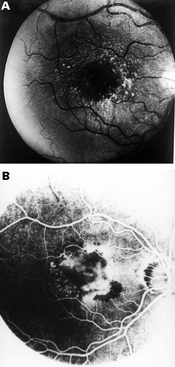
Figure 1 .

(A) Right eye. Fundus photograph in 1973 shows a radial pattern of innumerable small elongated basal laminar drusen. (B) Right eye. Fluorescein angiogram in 1981 shows submacular neovascularisation with fluorescein leakage.
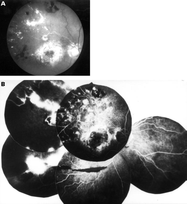
Figure 2 .
(A) Right eye. Photograph in 1996. Advanced stage of the macular disease with irregular subretinal fibrous metaplasia, hyperplasia of the retinal pigment epithelium, and discrete radial basal laminar drusen. Note also a wedge-shaped superotemporal area with intraretinal haemorrhages, hard exudates, and sheathed vessels. (B) Right eye. Fluorescein angiogram in 1996. Note the temporal retinal ischaemia with telangiectatic vessels, shunt vessels, and microaneurysms, in addition to the neovascularisation.


