Abstract
AIMS—To assess the clinical and fluorescein angiographic features of choroidal folds seen in association with papilloedema. METHODS—In a retrospective study, the clinical data from a database on patients with choroidal folds (1963-97), including fundus photography and fluorescein angiography, from 32 patients (64 eyes) with choroidal folds in association with papilloedema were reviewed. The clinical and fluorescein angiographic features and the clinical course of choroidal folds in these patients are described. RESULTS—32 patients had choroidal folds associated with papilloedema. Folds of two distinct categories were observed, either coarse folds or wrinkles. The folds persisted in all cases, even after resolution of papilloedema. Follow up ranged from 1 month to 20 years. Only one patient suffered permanent visual impairment as a result of a choroidal fold. CONCLUSIONS—Choroidal folds exist in two forms, coarse folds and wrinkles. They persist even after papilloedema has resolved. Final visual acuity did not appear to be affected by the presence of choroidal folds in the majority of patients.
Full Text
The Full Text of this article is available as a PDF (152.1 KB).
Figure 1 .
Visual loss due to macular folds. Colour photograph of the left fundus of a patient (patient no 8, Table 3) who developed papilloedema and choroidal folds as a result of benign intracranial hypertension. A heavily pigmented choroidal fold, which can be seen passing through the fovea, is responsible for permanent visual impairment. Visual acuity in the left eye is 2/60, and the patient describes severe left metamorphopsia.
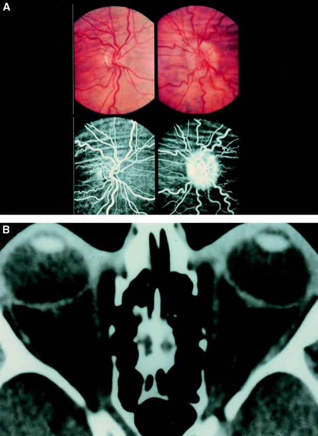
Figure 2 .

(A) Bilateral choroidal folds and unilateral papilloedema. Colour and fluorescein photographs demonstrating bilateral coarse choroidal folds and unilateral papilloedema in a patient (patient no 32, Table 3) with benign intracranial hypertension. (B) Distended nerve sheath and compressed globes. Computed tomograph scan of the same patient demonstrating dilated optic nerve sheaths. This patient presented with acquired hypermetropia. Examination revealed bilateral choroidal folds and a swollen left disc. The arrow denotes perineural cerebrospinal fluid.
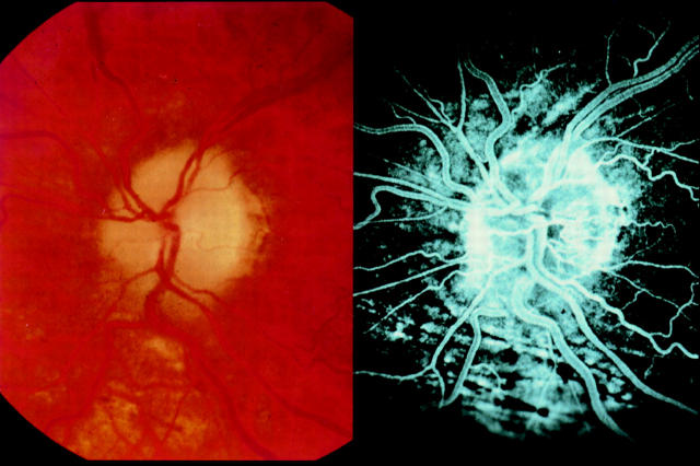
Figure 3 .
Subclinical choroidal folds. Colour fundus photography showing no evidence of choroidal folds in a patient with benign intracranial hypertension. Fluorescein angiogram of the same patient demonstrating the presence of "subclinical" choroidal folds.
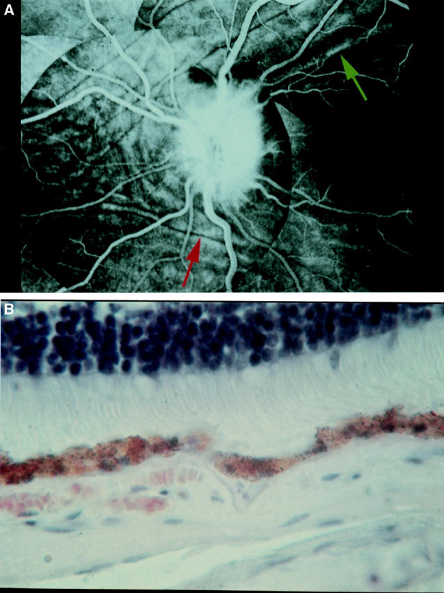
Figure 4 .

(A) Classic appearance of choroidal folds and papilloedema. This pattern of choroidal folding, where the folds curve around the nasal aspect of the disc, and then sweep superotemporally and inferotemporally, is the most common pattern of choroidal folding seen in association with papilloedema. Note the presence of both choroidal wrinkles (red arrow) and coarse choroidal folds (green arrow). (B) Histological section showing a wrinkle (arrow) involving only the retinal pigment epithelium and Bruch's membrane. The overlying retina is flat.
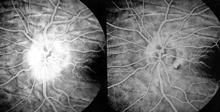
Figure 5 .
Persistence of choroidal folds. Fluorescein photographs showing the persistence of choroidal folds 7 years after resolution of papilloedema. This patient had a left optic nerve sheath fenestration for benign intracranial hypertension. Left: choroidal folds with papilloedema; right: 7 years after resolution.
Selected References
These references are in PubMed. This may not be the complete list of references from this article.
- Amalric P. Un nouveau tye de plis rétiniens congénitaux. Arch Ophtalmol Rev Gen Ophtalmol. 1968 Jul-Aug;28(5):507–512. [PubMed] [Google Scholar]
- Atta H. R., Byrne S. F. The findings of standardized echography for choroidal folds. Arch Ophthalmol. 1988 Sep;106(9):1234–1241. doi: 10.1001/archopht.1988.01060140394040. [DOI] [PubMed] [Google Scholar]
- Bird A. C., Sanders M. D. Choroidal folds in association with papilloedema. Br J Ophthalmol. 1973 Feb;57(2):89–97. doi: 10.1136/bjo.57.2.89. [DOI] [PMC free article] [PubMed] [Google Scholar]
- Bullock J. D., Egbert P. R. Experimental choroidal folds. Am J Ophthalmol. 1974 Oct;78(4):618–623. doi: 10.1016/s0002-9394(14)76299-3. [DOI] [PubMed] [Google Scholar]
- Cairns J. D. Disk oedema and choroidal folds. Aust J Ophthalmol. 1973 Feb;1(1):30–35. doi: 10.1111/j.1442-9071.1973.tb00106.x. [DOI] [PubMed] [Google Scholar]
- Cangemi F. E., Trempe C. L., Walsh J. B. Choroidal folds. Am J Ophthalmol. 1978 Sep;86(3):380–387. doi: 10.1016/0002-9394(78)90243-x. [DOI] [PubMed] [Google Scholar]
- Cappaert W. E., Purnell E. W., Frank K. E. Use of B-sector scan ultrasound in the diagnosis of benign choroidal folds. Am J Ophthalmol. 1977 Sep;84(3):375–379. doi: 10.1016/0002-9394(77)90682-1. [DOI] [PubMed] [Google Scholar]
- Gittinger J. W., Jr, Asdourian G. K. Macular abnormalities in papilledema from pseudotumor cerebri. Ophthalmology. 1989 Feb;96(2):192–194. doi: 10.1016/s0161-6420(89)32915-0. [DOI] [PubMed] [Google Scholar]
- Hayreh S. S. Optic disc edema in raised intracranial pressure. V. Pathogenesis. Arch Ophthalmol. 1977 Sep;95(9):1553–1565. doi: 10.1001/archopht.1977.04450090075006. [DOI] [PubMed] [Google Scholar]
- Hyvärinen L., Walsh F. B. Benign chorioretinal folds. Am J Ophthalmol. 1970 Jul;70(1):14–17. doi: 10.1016/0002-9394(70)90660-4. [DOI] [PubMed] [Google Scholar]
- Jacobson D. M. Intracranial hypertension and the syndrome of acquired hyperopia with choroidal folds. J Neuroophthalmol. 1995 Sep;15(3):178–185. [PubMed] [Google Scholar]
- Leahey A. B., Brucker A. J., Wyszynski R. E., Shaman P. Chorioretinal folds. A comparison of unilateral and bilateral cases. Arch Ophthalmol. 1993 Mar;111(3):357–359. doi: 10.1001/archopht.1993.01090030075042. [DOI] [PubMed] [Google Scholar]
- Liu D., Kahn M. Measurement and relationship of subarachnoid pressure of the optic nerve to intracranial pressures in fresh cadavers. Am J Ophthalmol. 1993 Nov 15;116(5):548–556. doi: 10.1016/s0002-9394(14)73195-2. [DOI] [PubMed] [Google Scholar]
- Newell F. W. Choroidal folds. The seventh Harry Searls Gradle Memorial lecture. Am J Ophthalmol. 1973 Jun;75(6):930–942. [PubMed] [Google Scholar]
- Norton E. W. A characteristic fluorescein angiographic pattern in choriodal folds. Proc R Soc Med. 1969 Feb;62(2):119–128. [PMC free article] [PubMed] [Google Scholar]
- SCHEPENS C. L., SCHWARTZ A. Intraocular tumors. I. Bilateral hemangioma of the choroid. AMA Arch Ophthalmol. 1958 Jul;60(1):72–83. [PubMed] [Google Scholar]
- Sanders M. D. The Bowman Lecture. Papilloedema: 'the pendulum of progress'. Eye (Lond) 1997;11(Pt 3):267–294. doi: 10.1038/eye.1997.63. [DOI] [PubMed] [Google Scholar]
- Wise G. N. Clinical features of idiopathic preretinal macular fibrosis. Schoenberg Lecture. Am J Ophthalmol. 1975 Mar;79(3):349–347. doi: 10.1016/0002-9394(75)90605-4. [DOI] [PubMed] [Google Scholar]
- von Winning C. H. Proceedings: Fluorography of choroidal folds. Ophthalmologica. 1973;167(5):436–439. doi: 10.1159/000306990. [DOI] [PubMed] [Google Scholar]


