Full Text
The Full Text of this article is available as a PDF (147.4 KB).
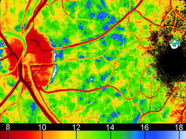
Figure 1 .

Parametric image in which the colour shows the time (in seconds) for the fluorescence to reach its maximum value. The patient has mild diabetic retinopathy.
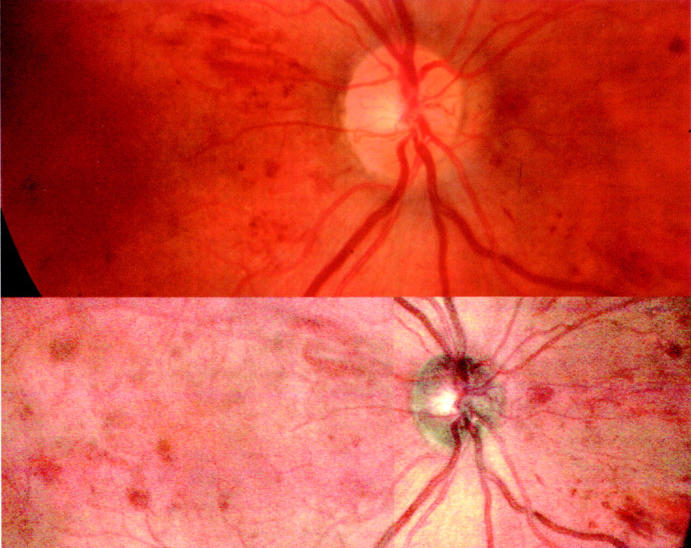
Figure 2 .

Upper image is a conventional fundus camera image of a patient with mild diabetic retinopathy. The lower image is the colour SLO picture of the same patient. Note that for the colour SLO image two separate images have been combined to give the requisite field of view.
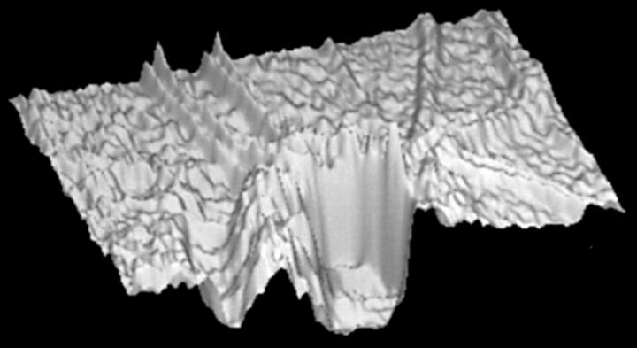
Figure 3 .
Three dimensional rendered topographic reconstruction of the retina through the optic disc.
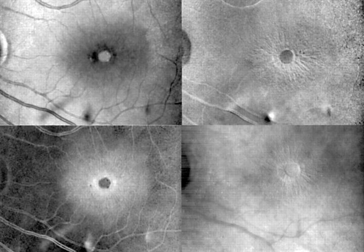
Figure 4 .
Patient with a macular hole. The top left is an SLO image of the fundus using an infrared laser; the top right shows a topographic image in which white indicates elevated areas; bottom left is a thickness image in which white indicates thicker areas; and bottom right is an indirect mode image.
Selected References
These references are in PubMed. This may not be the complete list of references from this article.
- Abadi R. V., Dickinson C. M. Monochromatic fundus photography of human albinos. Arch Ophthalmol. 1983 Nov;101(11):1706–1711. doi: 10.1001/archopht.1983.01040020708008. [DOI] [PubMed] [Google Scholar]
- Anton A., Zangwill L., Emdadi A., Weinreb R. N. Nerve fiber layer measurements with scanning laser polarimetry in ocular hypertension. Arch Ophthalmol. 1997 Mar;115(3):331–334. doi: 10.1001/archopht.1997.01100150333004. [DOI] [PubMed] [Google Scholar]
- Bartsch D. U., Freeman W. R. Axial intensity distribution analysis of the human retina with a confocal scanning laser tomograph. Exp Eye Res. 1994 Feb;58(2):161–173. doi: 10.1006/exer.1994.1004. [DOI] [PubMed] [Google Scholar]
- Bartsch D. U., Freeman W. R. Laser-tissue interaction and artifacts in confocal scanning laser ophthalmoscopy and tomography. Neurosci Biobehav Rev. 1993 Winter;17(4):459–467. doi: 10.1016/s0149-7634(05)80123-x. [DOI] [PubMed] [Google Scholar]
- Baumann M., Gentile R. C., Liebmann J. M., Ritch R. Reproducibility of retinal thickness measurements in normal eyes using optical coherence tomography. Ophthalmic Surg Lasers. 1998 Apr;29(4):280–285. [PubMed] [Google Scholar]
- Beausencourt E., Elsner A. E., Hartnett M. E., Trempe C. L. Quantitative analysis of macular holes with scanning laser tomography. Ophthalmology. 1997 Dec;104(12):2018–2029. doi: 10.1016/s0161-6420(97)30062-1. [DOI] [PubMed] [Google Scholar]
- Broadway D. C., Drance S. M., Parfitt C. M., Mikelberg F. S. The ability of scanning laser ophthalmoscopy to identify various glaucomatous optic disk appearances. Am J Ophthalmol. 1998 May;125(5):593–604. doi: 10.1016/s0002-9394(98)00002-6. [DOI] [PubMed] [Google Scholar]
- Bursell S. E., Clermont A. C., Kinsley B. T., Simonson D. C., Aiello L. M., Wolpert H. A. Retinal blood flow changes in patients with insulin-dependent diabetes mellitus and no diabetic retinopathy. Invest Ophthalmol Vis Sci. 1996 Apr;37(5):886–897. [PubMed] [Google Scholar]
- Cree M. J., Olson J. A., McHardy K. C., Sharp P. F., Forrester J. V. The preprocessing of retinal images for the detection of fluorescein leakage. Phys Med Biol. 1999 Jan;44(1):293–308. doi: 10.1088/0031-9155/44/1/021. [DOI] [PubMed] [Google Scholar]
- Delori F. C., Gragoudas E. S., Francisco R., Pruett R. C. Monochromatic ophthalmoscopy and fundus photography. The normal fundus. Arch Ophthalmol. 1977 May;95(5):861–868. doi: 10.1001/archopht.1977.04450050139018. [DOI] [PubMed] [Google Scholar]
- Ducrey N. M., Delori F. C., Gragoudas E. S. Monochromatic ophthalmoscopy and fundus photography. II. The pathological fundus. Arch Ophthalmol. 1979 Feb;97(2):288–293. doi: 10.1001/archopht.1979.01020010140009. [DOI] [PubMed] [Google Scholar]
- Freeman W. R., Bartsch D. U., Mueller A. J., Banker A. S., Weinreb R. N. Simultaneous indocyanine green and fluorescein angiography using a confocal scanning laser ophthalmoscope. Arch Ophthalmol. 1998 Apr;116(4):455–463. doi: 10.1001/archopht.116.4.455. [DOI] [PubMed] [Google Scholar]
- Goldbaum M. H., Katz N. P., Nelson M. R., Haff L. R. The discrimination of similarly colored objects in computer images of the ocular fundus. Invest Ophthalmol Vis Sci. 1990 Apr;31(4):617–623. [PubMed] [Google Scholar]
- Hammer M., Roggan A., Schweitzer D., Müller G. Optical properties of ocular fundus tissues--an in vitro study using the double-integrating-sphere technique and inverse Monte Carlo simulation. Phys Med Biol. 1995 Jun;40(6):963–978. doi: 10.1088/0031-9155/40/6/001. [DOI] [PubMed] [Google Scholar]
- Haynes R. J., Manivannan A., Walker S., Sharp P. F., Forrester J. V. Imaging of optic nerve head drusen with the scanning laser ophthalmoscope. Br J Ophthalmol. 1997 Aug;81(8):654–657. doi: 10.1136/bjo.81.8.654. [DOI] [PMC free article] [PubMed] [Google Scholar]
- Hee M. R., Baumal C. R., Puliafito C. A., Duker J. S., Reichel E., Wilkins J. R., Coker J. G., Schuman J. S., Swanson E. A., Fujimoto J. G. Optical coherence tomography of age-related macular degeneration and choroidal neovascularization. Ophthalmology. 1996 Aug;103(8):1260–1270. doi: 10.1016/s0161-6420(96)30512-5. [DOI] [PubMed] [Google Scholar]
- Hee M. R., Puliafito C. A., Duker J. S., Reichel E., Coker J. G., Wilkins J. R., Schuman J. S., Swanson E. A., Fujimoto J. G. Topography of diabetic macular edema with optical coherence tomography. Ophthalmology. 1998 Feb;105(2):360–370. doi: 10.1016/s0161-6420(98)93601-6. [DOI] [PMC free article] [PubMed] [Google Scholar]
- Hipwell J. H., Manivannan A., Vieira P., Sharp P. F., Forrester J. V. Quantifying changes in retinal circulation: the generation of parametric images from fluorescein angiograms. Physiol Meas. 1998 May;19(2):165–180. doi: 10.1088/0967-3334/19/2/004. [DOI] [PubMed] [Google Scholar]
- Hossain P., Liversidge J., Cree M. J., Manivannan A., Vieira P., Sharp P. F., Brown G. C., Forrester J. V. In vivo cell tracking by scanning laser ophthalmoscopy: quantification of leukocyte kinetics. Invest Ophthalmol Vis Sci. 1998 Sep;39(10):1879–1887. [PubMed] [Google Scholar]
- Howe L. J., Stanford M. R., Whiston R., Dewhirst R., Marshall J. Angiographic abnormalities of experimental autoimmune uveoretinitis. Curr Eye Res. 1996 Dec;15(12):1149–1155. doi: 10.3109/02713689608995149. [DOI] [PubMed] [Google Scholar]
- Hudson C., Flanagan J. G., Turner G. S., McLeod D. Scanning laser tomography Z profile signal width as an objective index of macular retinal thickening. Br J Ophthalmol. 1998 Feb;82(2):121–130. doi: 10.1136/bjo.82.2.121. [DOI] [PMC free article] [PubMed] [Google Scholar]
- Ikeda T., Sato K., Katano T., Hayashi Y. Examination of patients with cystoid macular edema using a scanning laser ophthalmoscope with infrared light. Am J Ophthalmol. 1998 May;125(5):710–712. doi: 10.1016/s0002-9394(98)00008-7. [DOI] [PubMed] [Google Scholar]
- Ikeda T., Sato K., Katano T., Hayashi Y. Examination of patients with cystoid macular edema using a scanning laser ophthalmoscope with infrared light. Am J Ophthalmol. 1998 May;125(5):710–712. doi: 10.1016/s0002-9394(98)00008-7. [DOI] [PubMed] [Google Scholar]
- Irak I., Zangwill L., Garden V., Shakiba S., Weinreb R. N. Change in optic disk topography after trabeculectomy. Am J Ophthalmol. 1996 Nov;122(5):690–695. doi: 10.1016/s0002-9394(14)70488-x. [DOI] [PubMed] [Google Scholar]
- Junghardt A., Schmid M. K., Schipper I., Wildberger H., Seifert B. Reproducibility of the data determined by scanning laser polarimetry. Graefes Arch Clin Exp Ophthalmol. 1996 Oct;234(10):628–632. doi: 10.1007/BF00185296. [DOI] [PubMed] [Google Scholar]
- Kaefer O. Degenerative changes in retinal vessels. Photodocumentation with monochromatic filters. Arch Ophthalmol. 1980 Feb;98(2):303–306. doi: 10.1001/archopht.1980.01020030299012. [DOI] [PubMed] [Google Scholar]
- Kinukawa Y., Shimura M., Tamai M. Quantifying leukocyte dynamics and plugging in retinal microcirculation of streptozotosin-induced diabetic rats. Curr Eye Res. 1999 Jan;18(1):49–55. doi: 10.1076/ceyr.18.1.49.5389. [DOI] [PubMed] [Google Scholar]
- Kirkpatrick J. N., Manivannan A., Gupta A. K., Hipwell J., Forrester J. V., Sharp P. F. Fundus imaging in patients with cataract: role for a variable wavelength scanning laser ophthalmoscope. Br J Ophthalmol. 1995 Oct;79(10):892–899. doi: 10.1136/bjo.79.10.892. [DOI] [PMC free article] [PubMed] [Google Scholar]
- Lachkar Y., Cohn H. Reproducibility of optic nerve head topographic measurements with the glaucoma-scope. Eye (Lond) 1997;11(Pt 6):810–817. doi: 10.1038/eye.1997.211. [DOI] [PubMed] [Google Scholar]
- Manivannan A., Kirkpatrick J. N., Sharp P. F., Forrester J. V. Novel approach towards colour imaging using a scanning laser ophthalmoscope. Br J Ophthalmol. 1998 Apr;82(4):342–345. doi: 10.1136/bjo.82.4.342. [DOI] [PMC free article] [PubMed] [Google Scholar]
- Mulholland D. A., Craig J. J., Rankin S. J. Use of scanning laser ophthalmoscopy to monitor papilloedema in idiopathic intracranial hypertension. Br J Ophthalmol. 1998 Nov;82(11):1301–1305. doi: 10.1136/bjo.82.11.1301. [DOI] [PMC free article] [PubMed] [Google Scholar]
- Mulholland D. A., Craig J. J., Rankin S. J. Use of scanning laser ophthalmoscopy to monitor papilloedema in idiopathic intracranial hypertension. Br J Ophthalmol. 1998 Nov;82(11):1301–1305. doi: 10.1136/bjo.82.11.1301. [DOI] [PMC free article] [PubMed] [Google Scholar]
- Orgül S., Cioffi G. A., Bacon D. R., Van Buskirk E. M. Sources of variability of topometric data with a scanning laser ophthalmoscope. Arch Ophthalmol. 1996 Feb;114(2):161–164. doi: 10.1001/archopht.1996.01100130155007. [DOI] [PubMed] [Google Scholar]
- Phillips R. P., Spencer T., Ross P. G., Sharp P. F., Forrester J. V. Quantification of diabetic maculopathy by digital imaging of the fundus. Eye (Lond) 1991;5(Pt 1):130–137. doi: 10.1038/eye.1991.24. [DOI] [PubMed] [Google Scholar]
- Poinoosawmy D., Fontana L., Wu J. X., Fitzke F. W., Hitchings R. A. Variation of nerve fibre layer thickness measurements with age and ethnicity by scanning laser polarimetry. Br J Ophthalmol. 1997 May;81(5):350–354. doi: 10.1136/bjo.81.5.350. [DOI] [PMC free article] [PubMed] [Google Scholar]
- Puliafito C. A., Hee M. R., Lin C. P., Reichel E., Schuman J. S., Duker J. S., Izatt J. A., Swanson E. A., Fujimoto J. G. Imaging of macular diseases with optical coherence tomography. Ophthalmology. 1995 Feb;102(2):217–229. doi: 10.1016/s0161-6420(95)31032-9. [DOI] [PubMed] [Google Scholar]
- Remky A., Beausencourt E., Elsner A. E. Angioscotometry with the scanning laser ophthalmoscope. Comparison of the effect of different wavelengths. Invest Ophthalmol Vis Sci. 1996 Oct;37(11):2350–2355. [PubMed] [Google Scholar]
- Roh S., Noecker R. J., Schuman J. S., Hedges T. R., 3rd, Weiter J. J., Mattox C. Effect of optic nerve head drusen on nerve fiber layer thickness. Ophthalmology. 1998 May;105(5):878–885. doi: 10.1016/S0161-6420(98)95031-X. [DOI] [PMC free article] [PubMed] [Google Scholar]
- Rutledge B. K., Puliafito C. A., Duker J. S., Hee M. R., Cox M. S. Optical coherence tomography of macular lesions associated with optic nerve head pits. Ophthalmology. 1996 Jul;103(7):1047–1053. doi: 10.1016/s0161-6420(96)30568-x. [DOI] [PubMed] [Google Scholar]
- Schneider U., Sherif-Adel S., Gelisken F., Inhoffen W., Kreissig I. Detection of choroidal aneurysms with indocyanine green videoangiography. Graefes Arch Clin Exp Ophthalmol. 1998 Mar;236(3):193–195. doi: 10.1007/s004170050063. [DOI] [PubMed] [Google Scholar]
- Schuman J. S., Pedut-Kloizman T., Hertzmark E., Hee M. R., Wilkins J. R., Coker J. G., Puliafito C. A., Fujimoto J. G., Swanson E. A. Reproducibility of nerve fiber layer thickness measurements using optical coherence tomography. Ophthalmology. 1996 Nov;103(11):1889–1898. doi: 10.1016/s0161-6420(96)30410-7. [DOI] [PMC free article] [PubMed] [Google Scholar]
- Schweitzer D., Hammer M., Scibor M. Imaging spectrometry in ophthalmology--principle and applications in microcirculation and in investigation of pigments. Ophthalmic Res. 1996;28 (Suppl 2):37–44. doi: 10.1159/000267955. [DOI] [PubMed] [Google Scholar]
- Sharp P. F., Manivannan A. The scanning laser ophthalmoscope. Phys Med Biol. 1997 May;42(5):951–966. doi: 10.1088/0031-9155/42/5/014. [DOI] [PubMed] [Google Scholar]
- Vieira P., Manivannan A., Lim C. S., Sharp P., Forrester J. V. Tomographic reconstruction of the retina using a confocal scanning laser ophthalmoscope. Physiol Meas. 1999 Feb;20(1):1–19. doi: 10.1088/0967-3334/20/1/001. [DOI] [PubMed] [Google Scholar]
- Ward N. P., Tomlinson S., Taylor C. J. Image analysis of fundus photographs. The detection and measurement of exudates associated with diabetic retinopathy. Ophthalmology. 1989 Jan;96(1):80–86. [PubMed] [Google Scholar]
- Weinreb R. N., Zangwill L., Berry C. C., Bathija R., Sample P. A. Detection of glaucoma with scanning laser polarimetry. Arch Ophthalmol. 1998 Dec;116(12):1583–1589. doi: 10.1001/archopht.116.12.1583. [DOI] [PubMed] [Google Scholar]
- Zambarakji H. J., Evans J. E., Amoaku W. M., Vernon S. A. Reproducibility of volumetric measurements of normal maculae with the Heidelberg retina tomograph. Br J Ophthalmol. 1998 Aug;82(8):884–891. doi: 10.1136/bjo.82.8.884. [DOI] [PMC free article] [PubMed] [Google Scholar]
- Zangwill L., Berry C. A., Garden V. S., Weinreb R. N. Reproducibility of retardation measurements with the nerve fiber analyzer II. J Glaucoma. 1997 Dec;6(6):384–389. [PubMed] [Google Scholar]
- van Norren D., van de Kraats J. Imaging retinal densitometry with a confocal Scanning Laser Ophthalmoscope. Vision Res. 1989;29(12):1825–1830. doi: 10.1016/0042-6989(89)90163-6. [DOI] [PubMed] [Google Scholar]
- von Rückmann A., Fitzke F. W., Bird A. C. Distribution of fundus autofluorescence with a scanning laser ophthalmoscope. Br J Ophthalmol. 1995 May;79(5):407–412. doi: 10.1136/bjo.79.5.407. [DOI] [PMC free article] [PubMed] [Google Scholar]
- von Rückmann A., Fitzke F. W., Bird A. C. In vivo fundus autofluorescence in macular dystrophies. Arch Ophthalmol. 1997 May;115(5):609–615. doi: 10.1001/archopht.1997.01100150611006. [DOI] [PubMed] [Google Scholar]


