Abstract
AIMS—To investigate the histopathological changes of rabbit corneas after laser in situ keratomileusis (LASIK) and to evaluate the corneal wound healing process. METHODS—A LASIK was performed on white rabbit eyes. Postoperatively, rabbits were killed on days 1 and 7, and at 1, 3, and 9 months. RESULTS—Periodic acid Schiff (PAS) positive material and disorganised collagen fibre were seen along the interface of the corneal flap even 9 months after operation. CONCLUSIONS—The wound healing process still continued at 9 months after LASIK indicating that a much longer time than expected was required for corneal wound healing following LASIK.
Full Text
The Full Text of this article is available as a PDF (104.6 KB).
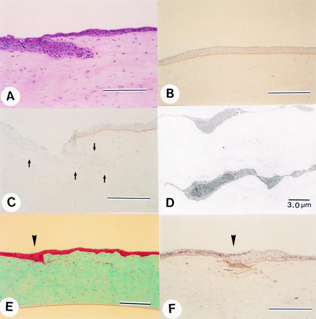
Figure 1 .
(A) Haematoxylin and eosin staining section on day 1 after LASIK. (bar 200 µm). (B) Normal epithelial basement membrane structure is observed at the centre of the cornea. (Anti-type IV collagen, postoperative day 1, bar 200 µm.) (C) At the wound margin, epithelial basement membrane structure is absent (arrows). (Anti-type IV collagen, postoperative day 1, bar 200 µm). (D) Electron microscopy showing wavy keratocyte with swelling the stroma in the corneal flap (postoperative day 1). (E) One week after LASIK. Masson trichrome staining section shows the epithelial plug is smaller than on day 1 after the surgery. Arrowhead indicates the wound edge (bar 200 µm). (F) Diffuse staining for type IV collagen is noted beneath the epithelium at the wound margin. Arrowhead indicates the wound edge (postoperative day 7, bar 200 µm).
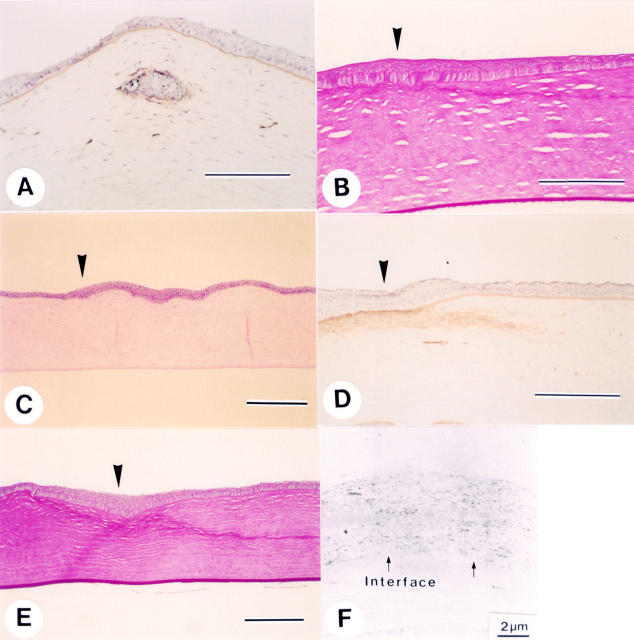
Figure 2 .
(A) Positive staining for type IV collagen is seen around the epithelial ingrowth portion on day 7 after LASIK (bar 200 µm). (B) PAS positive material is noted along the lamellar interface. Arrowhead indicates the wound edge (postoperative month 1, bar 200 µm). (C) Haematoxylin and eosin staining section 3 months after LASIK showing almost normal appearing cornea. Arrowhead indicates the wound edge (bar 200 µm). (D) Diffuse staining for type IV collagen is seen beneath the epithelium at the wound margin. Arrowhead indicates the wound edge (postoperative 3 months, bar 200 µm). (E) PAS stained section 9 months after LASIK. Note that the PAS positive material still present along the interface. Arrowhead indicates the wound edge (bar 200 µm). (F) Electron microscopy at 9 months after LASIK. Disorganised extracellular matrix is deposited along the interface (arrows).
Selected References
These references are in PubMed. This may not be the complete list of references from this article.
- Amm M., Wetzel W., Winter M., Uthoff D., Duncker G. I. Histopathological comparison of photorefractive keratectomy and laser in situ keratomileusis in rabbits. J Refract Surg. 1996 Nov-Dec;12(7):758–766. doi: 10.3928/1081-597X-19961101-07. [DOI] [PubMed] [Google Scholar]
- Buratto L., Ferrari M., Rama P. Excimer laser intrastromal keratomileusis. Am J Ophthalmol. 1992 Mar 15;113(3):291–295. doi: 10.1016/s0002-9394(14)71581-8. [DOI] [PubMed] [Google Scholar]
- Güell J. L., Muller A. Laser in situ keratomileusis (LASIK) for myopia from -7 to -18 diopters. J Refract Surg. 1996 Feb;12(2):222–228. doi: 10.3928/1081-597X-19960201-03. [DOI] [PubMed] [Google Scholar]
- Helena M. C., Baerveldt F., Kim W. J., Wilson S. E. Keratocyte apoptosis after corneal surgery. Invest Ophthalmol Vis Sci. 1998 Feb;39(2):276–283. [PubMed] [Google Scholar]
- Helena M. C., Meisler D., Wilson S. E. Epithelial growth within the lamellar interface after laser in situ keratomileusis (LASIK) Cornea. 1997 May;16(3):300–305. [PubMed] [Google Scholar]
- Kato T., Nakayasu K., Ikegami K., Obara T., Kanayama T., Kanai A. Analysis of glycosaminoglycans in rabbit cornea after excimer laser keratectomy. Br J Ophthalmol. 1999 May;83(5):609–612. doi: 10.1136/bjo.83.5.609. [DOI] [PMC free article] [PubMed] [Google Scholar]
- Kato T., Watanabe Y., Nakayasu K., Kanai A., Yajima Y. The ultrastructure of the lens capsule abnormalities in Alport's syndrome. Jpn J Ophthalmol. 1998 Sep-Oct;42(5):401–405. doi: 10.1016/s0021-5155(98)00031-8. [DOI] [PubMed] [Google Scholar]
- Maldonado-Bas A., Onnis R. Results of laser in situ keratomileusis in different degrees of myopia. Ophthalmology. 1998 Apr;105(4):606–611. doi: 10.1016/S0161-6420(98)94012-X. [DOI] [PubMed] [Google Scholar]
- Pallikaris I. G., Papatzanaki M. E., Stathi E. Z., Frenschock O., Georgiadis A. Laser in situ keratomileusis. Lasers Surg Med. 1990;10(5):463–468. doi: 10.1002/lsm.1900100511. [DOI] [PubMed] [Google Scholar]
- Pérez-Santonja J. J., Bellot J., Claramonte P., Ismail M. M., Alió J. L. Laser in situ keratomileusis to correct high myopia. J Cataract Refract Surg. 1997 Apr;23(3):372–385. doi: 10.1016/s0886-3350(97)80182-4. [DOI] [PubMed] [Google Scholar]
- Pérez-Santonja J. J., Linna T. U., Tervo K. M., Sakla H. F., Alió y Sanz J. L., Tervo T. M. Corneal wound healing after laser in situ keratomileusis in rabbits. J Refract Surg. 1998 Nov-Dec;14(6):602–609. doi: 10.3928/1081-597X-19981101-06. [DOI] [PubMed] [Google Scholar]
- Salah T., Waring G. O., 3rd, el Maghraby A., Moadel K., Grimm S. B. Excimer laser in situ keratomileusis under a corneal flap for myopia of 2 to 20 diopters. Am J Ophthalmol. 1996 Feb;121(2):143–155. [PubMed] [Google Scholar]


