

Abstract
AIM—To determine if the isoforms of transforming growth factor β (TGF-β) are present in fetal, normal adult, and glaucomatous optic nerve heads. METHODS—To localise cells synthesising TGF-β, optic nerve heads were stained using antibodies to TGF-β1, TGF-β2, and TGF-β3. To demonstrate synthesis, human optic nerve heads from fetal, glaucomatous, and normal age matched subjects were explanted, cultured overnight, and the culture supernatant was assayed for the presence of TGF-β1 and TGF-β2 by bioassay. In addition, semiquantitative RT-PCR was performed to determine the gene expression pattern of TGF-β2. RESULTS—Immunohistochemistry of glaucomatous samples revealed the presence of intense staining for TGF-β2 primarily in astrocytes, whereas TGF-β1 was localised to blood vessels. No TGF-β3 immunoreactivity was observed. There was little or no expression of TGF-β in normal optic nerve heads. Optic nerve heads from glaucomatous eyes released 70-100-fold more TGF-β2 than normal age matched optic nerve heads. Fetal optic nerve heads released 90-100-fold more TGF-β2 than normal adult optic nerve heads. TGF-β1 was undetectable by bioassay in all samples tested. There was no apparent increase in TGF-β2 gene expression in glaucomatous and fetal eyes, suggesting post-transcriptional regulatory mechanisms. CONCLUSIONS—These results demonstrate that TGF-β2 is produced in high levels in the fetal and glaucomatous optic nerve heads, perhaps by a mechanism of post-transcriptional regulation. TGF-β may be important during development of the optic nerve head and, in glaucoma, TGF-β2 may be a mediator of astrocyte reactivation and extracellular matrix remodelling in the lamina cribrosa. Keywords: astrocytes; glaucoma; optic nerve head; transforming growth factor β
Full Text
The Full Text of this article is available as a PDF (275.0 KB).
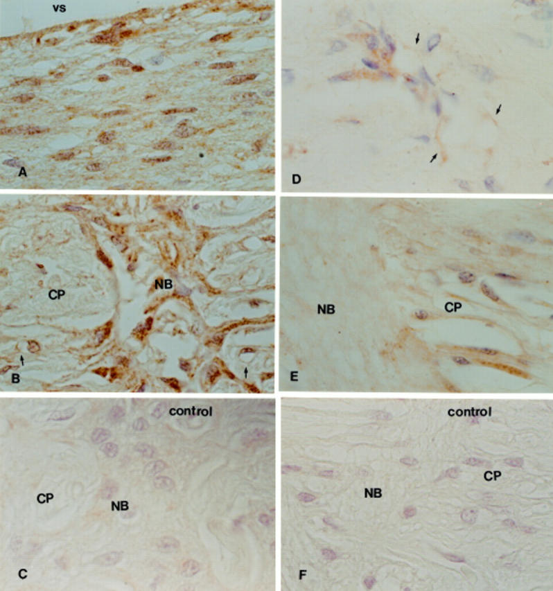
Figure 1 .

TGF-β1 and TGF-β2 immunoperoxidase staining in normal optic nerve head, donor age 68 years old. Little or no staining for TGF-β2 is observed. Few blood vessels were stained with TGF-β1 (arrow, E). (A) and (D) Prelaminar region; (B) and (E) lamina cribrosa; (C) and (F) negative controls. vs = vitreal surface; CP = cribriform plates, NB = nerve bundles. ×375.
Figure 2 .
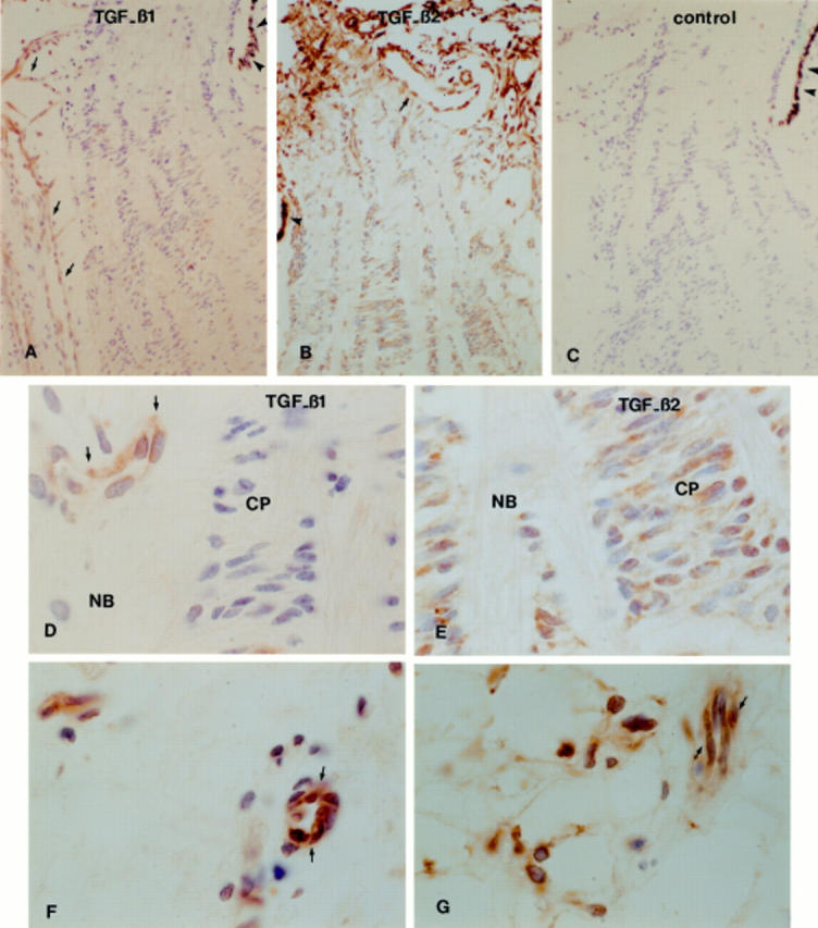
TGF-β1 and TGF-β2 immunoperoxidase staining in fetal optic nerve head (gestational age 24 weeks). TGF-β1 immunoreactivity was restricted to blood vessels throughout the optic nerve head (A, D) and TGF-β2 reactivity was localised to the extracellular matrix and cells in the optic nerve head (B, E). In the sclera, TGF-β1 was detected in association with blood vessels (F) and TGF-β2 was associated with the vessels and fibroblasts (G). (C) Illustrates the negative control. Arrows point to blood vessels and arrowheads point to the retinal pigment epithelium. NB = nerve bundles; CP = cribriform plates. (A-C) ×75; (D-G) ×750.
Figure 3 .

TGF-β1 immunoperoxidase staining in advanced primary open angle glaucoma, donor age 78 years old. Note staining around blood vessels (arrows). (A) Prelaminar region; (B) lamina cribrosa; (C) negative control. vs = vitreal surface; CP = cribriform plates; NB = nerve bundles. ×350.
Figure 4 .
TGF-β2 immunoperoxidase staining in advanced (A, B, and C) and mild (D, E, and F) primary open angle glaucoma, donor ages 74 years old (advanced glaucoma) and 67 years old (mild glaucoma). Note widespread staining of astrocytes and the extracellular matrix throughout the optic nerve head of the sample from with advanced glaucoma (A and B). Reactivity in blood vessels was also noted (arrow). In the mild glaucoma sample, few astrocytes and blood vessels (arrows) were labelled in the prelaminar region (D) and in the lamina cribrosa, with little staining in the extracellular matrix (E). (C) and (F) are negative controls. vs = vitreal surface; NB = nerve bundles; CP = cribriform plates. ×750.
Figure 5 .

Co-localisation of TGF-β2 and GFAP by immunofluorescence in the lamina cribrosa of a glaucomatous donor (age 92 years old). In (A) immunoreactivity for TGF-β2 is localised to cells inside the cribriform plates (CP) and in the nerve bundles (NB). These cells are also GFAP positive, confirming their identity as astrocytes (B). (C) Illustrates the co-localisation of TGF-β2 and GFAP. Arrows point to some of the cells that show double staining. ×400.
Figure 6 .

TGF-β2 mRNA levels in fetal, normal adult, and glaucomatous eyes, as determined by relative RT-PCR. Similar levels of TGF-β2 mRNA were observed in all samples tested (upper), when compared with β actin levels (lower). Lane 1, molecular weight marker (100 bp ladder, FMC Products); lane 2, cultured human brain astrocytes; lane 3, cultured type 1B astrocytes; lanes 4 and 5, fetal optic nerve heads, 19 and 20 weeks gestational age; lanes 6-8, normal adult optic nerve heads, donor ages 66, 88, and 94 years old; lanes 9-11, glaucomatous optic nerve heads, donor ages 53, 67, and 92 years old.