Abstract
BACKGROUND—Ring melanoma of the ciliary body and iris is extremely rare and often has adverse histology. This tumour may cause raised intraocular pressure. METHODS—A review of four cases of ring melanomas with insidious presentations seen in the ocular oncology service over a 12 month period. RESULTS—All four patients presented with unilateral anterior segment abnormalities and refractory glaucoma. The misdiagnoses of the causes of the glaucoma included angle recession from previous blunt trauma (patient 1); iridocorneal endothelial (ICE) syndrome supported by endothelial specular microscopy (patients 2 and 3); and melanocytoma on ciliary body biopsy (patient 4). Two patients were treated by several cyclodiode ciliary body ablation treatments and the other two underwent trabeculectomies and Molteno tubes. Two of the four patients have since died from their disease. CONCLUSION—The ophthalmologist should re-evaluate the diagnosis in patients with anterior segment abnormalities and refractory ipsilateral glaucoma. Endothelial specular microscopy and biopsy of the suspicious lesion may give misleading reassurance. The potential presence of an anterior uveal melanoma must always be considered. Keywords: ring melanoma; iridocorneal endothelial syndrome; glaucoma; uveal melanoma
Full Text
The Full Text of this article is available as a PDF (136.2 KB).
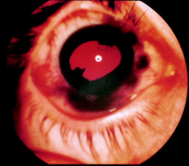
Figure 1 .
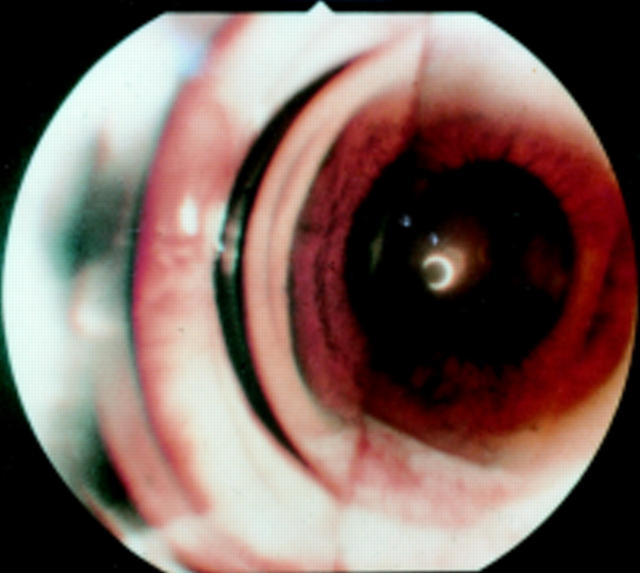
Circumcorneal dark shadow of ring melanoma.
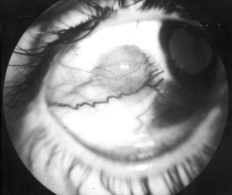
Figure 2 .
Conjunctival nodule of extrascleral extension of ring melanoma.
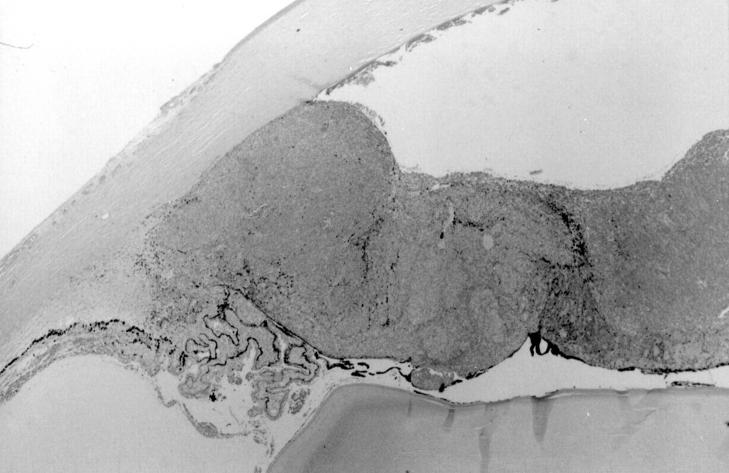
Figure 3 .
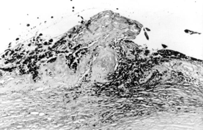
Low power view of the iris in the enucleation specimen showing an extensive melanocytic lesion filling the angle, invading the trabecular meshwork, and replacing the iris. (Haematoxylin and eosin, original magnification, ×20).
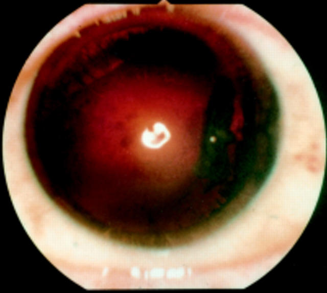
Figure 4 .
Focal lens opacity.
Figure 5 .
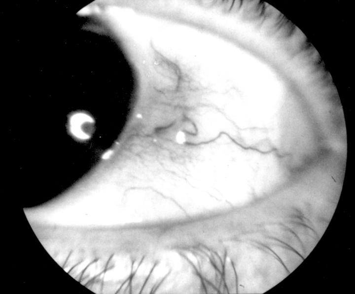
Sentinel vessel over pigmented iridociliary lesion.
Figure 6 .
Darkly pigmented angle on gonioscopy.
Figure 7 .
Obliteration of the ciliary processes by cyclodiode therapy adjacent to an area of infiltration of the root of the iris and trabecular meshwork by densely pigmented cells (periodic acid Schiff, original magnification, ×100).
Selected References
These references are in PubMed. This may not be the complete list of references from this article.
- Augsburger J. J., Affel L. L., Benarosh D. A. Ultrasound biomicroscopy of cystic lesions of the iris and ciliary body. Trans Am Ophthalmol Soc. 1996;94:259–274. doi: 10.1016/s0002-9394(14)70165-5. [DOI] [PMC free article] [PubMed] [Google Scholar]
- Barr C. C., McLean I. W., Zimmerman L. E. Uveal melanoma in children and adolescents. Arch Ophthalmol. 1981 Dec;99(12):2133–2136. doi: 10.1001/archopht.1981.03930021009003. [DOI] [PubMed] [Google Scholar]
- Brown D., Boniuk M., Font R. L. Diffuse malignant melanoma of iris with metastases. Surv Ophthalmol. 1990 Mar-Apr;34(5):357–364. doi: 10.1016/0039-6257(90)90111-8. [DOI] [PubMed] [Google Scholar]
- Chaudhry I. M., Moster M. R., Augsburger J. J. Iris ring melanoma masquerading as pigmentary glaucoma. Arch Ophthalmol. 1997 Nov;115(11):1480–1481. doi: 10.1001/archopht.1997.01100160650029. [DOI] [PubMed] [Google Scholar]
- Fitzpatrick M., Augsburger J. J., Koreishi F. M., Eagle R. C., Jr Complete ring melanoma of the choroid. Retina. 1996;16(3):228–231. doi: 10.1097/00006982-199616030-00008. [DOI] [PubMed] [Google Scholar]
- Font R. L., Spaulding A. G., Zimmerman LE DIFFUSE MALI a clinicopathologic report of 54 cases. Trans Am Acad Ophthalmol Otolaryngol. 1968 Nov-Dec;72(6):877–895. [PubMed] [Google Scholar]
- Foos R. Y., Hull S. N., Straatsma B. R. Early diagnosis of ciliary body melanomas. Arch Ophthalmol. 1969 Mar;81(3):336–344. doi: 10.1001/archopht.1969.00990010338007. [DOI] [PubMed] [Google Scholar]
- Frangoulis M. A., Sherrard E. S., Muir M. G., Buckley R. J. Clinical features of the irido-corneal endothelial syndrome. Trans Ophthalmol Soc U K. 1985;104(Pt 7):775–781. [PubMed] [Google Scholar]
- Jensen O. A., Johansen S., Kiss K. Intraocular T-cell lymphoma mimicking a ring melanoma. First manifestation of systemic disease. Report of a case and survey of the literature. Graefes Arch Clin Exp Ophthalmol. 1994 Mar;232(3):148–152. doi: 10.1007/BF00176784. [DOI] [PubMed] [Google Scholar]
- KURZ G. H. MALIGNANT MELANOMA OF CILIARY BODY AND IRIS MANIFESTED IN TWO LOCATIONS. Am J Ophthalmol. 1965 May;59:917–921. [PubMed] [Google Scholar]
- Kremer I., Ben-Sira I. Borderline spindle cell nevus of the ciliary body as a second primary melanoma--a clinicopathologic case. Ann Ophthalmol. 1989 Jun;21(6):217–221. [PubMed] [Google Scholar]
- Levy S. G., Kirkness C. M., Moss J., Ficker L., McCartney A. C. On the pathology of the iridocorneal-endothelial syndrome: the ultrastructural appearances of 'subtotal-ice'. Eye (Lond) 1995;9(Pt 3):318–323. doi: 10.1038/eye.1995.62. [DOI] [PubMed] [Google Scholar]
- Omulecki W., Pruszczyński M., Borowski J. Ring melanoma of the iris and ciliary body. Br J Ophthalmol. 1985 Jul;69(7):514–518. doi: 10.1136/bjo.69.7.514. [DOI] [PMC free article] [PubMed] [Google Scholar]
- RONES B., ZIMMERMAN L. E. The production of heterochromia and glaucoma by diffuse malignant melanoma of the iris. Trans Am Acad Ophthalmol Otolaryngol. 1957 Jul-Aug;61(4):447–463. [PubMed] [Google Scholar]
- Reese A. B., Howard G. M. Flat uveal melanomas. Am J Ophthalmol. 1967 Dec;64(6):1021–1028. doi: 10.1016/0002-9394(67)93054-1. [DOI] [PubMed] [Google Scholar]
- Shields C. L., Shields J. A., Shields M. B., Augsburger J. J. Prevalence and mechanisms of secondary intraocular pressure elevation in eyes with intraocular tumors. Ophthalmology. 1987 Jul;94(7):839–846. doi: 10.1016/s0161-6420(87)33537-7. [DOI] [PubMed] [Google Scholar]
- Shields M. B., Klintworth G. K. Anterior uveal melanomas and intraocular pressure. Ophthalmology. 1980 Jun;87(6):503–517. doi: 10.1016/s0161-6420(80)35202-0. [DOI] [PubMed] [Google Scholar]
- Singer P. R., Krupin T., Smith M. E., Becker B. Recurrent orbital and metastatic melanoma in a patient undergoing previous glaucoma surgery. Am J Ophthalmol. 1979 Jun;87(6):766–768. doi: 10.1016/0002-9394(79)90350-7. [DOI] [PubMed] [Google Scholar]
- Spaulding A. G., Green W. R., Font R. L. Ring-shaped limbal tumor. Secondary to unrecognized diffuse malignant melanoma of the uvea. Arch Ophthalmol. 1967 Jan;77(1):76–80. doi: 10.1001/archopht.1967.00980020078016. [DOI] [PubMed] [Google Scholar]


