Abstract
BACKGROUND—Iris cysts in children are uncommon and there is relatively little information on their classification, incidence, and management. METHODS—The records of all children under age 20 years who were diagnosed with iris cyst were reviewed and the types and incidence of iris cysts of childhood determined. Based on these observations recommendations were made regarding management of iris cysts in children. RESULTS—Of 57 iris cysts in children, 53 were primary and four were secondary. There were 44 primary cysts of the iris pigment epithelium, 34 of which were of the peripheral or iridociliary type, accounting for 59% of all childhood iris cysts. It was most commonly diagnosed in the teenage years, more common in girls (68%), was not recognised in infancy, remained stationary or regressed, and required no treatment. The five mid-zonal pigment epithelial cysts were diagnosed at a mean age of 14 years, were more common in boys (83%), remained stationary, and required no treatment. The pupillary type of pigment epithelial cyst was generally recognised in infancy and, despite involvement of the pupillary aperture, also required no treatment. There were nine cases of primary iris stromal cysts, accounting for 16% of all childhood iris cysts. This cyst was usually diagnosed in infancy, was generally progressive, and required treatment in eight of the nine cases, usually by aspiration and cryotherapy or surgical resection. Among the secondary iris cysts, two were post-traumatic epithelial ingrowth cysts and two were tumour induced cysts, one arising from an intraocular lacrimal gland choristoma and one adjacent to a peripheral iris naevus. CONCLUSIONS—Most iris cysts of childhood are primary pigment epithelial cysts and require no treatment. However, the iris stromal cyst, usually recognised in infancy, is generally an aggressive lesion that requires treatment by aspiration or surgical excision. Keywords: iris cysts; children
Full Text
The Full Text of this article is available as a PDF (133.0 KB).
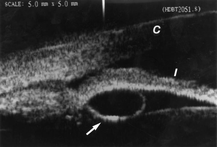
Figure 1 .
Peripheral iris pigment epithelial cyst. Ultrasound biomicroscopy, showing clear cyst (arrow) posterior to the iris (I). The cornea (C) is towards the top of the photograph.
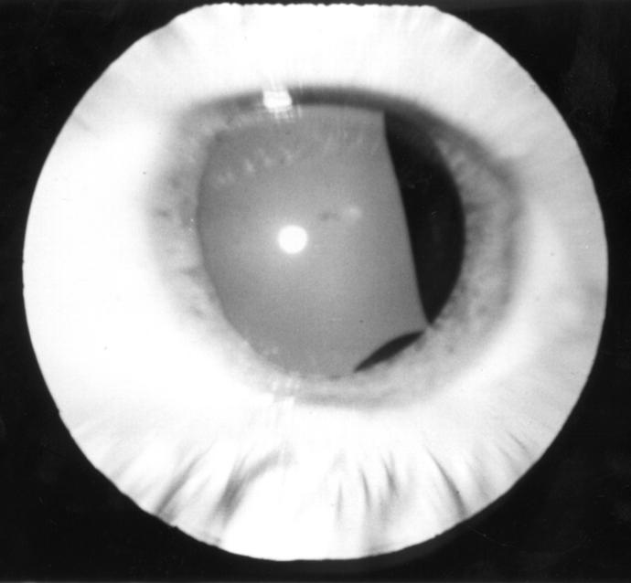
Figure 2 .
Mid-zonal iris pigment epithelial cyst. Note that there are two cysts, each of which has an elongated, fusiform shape.
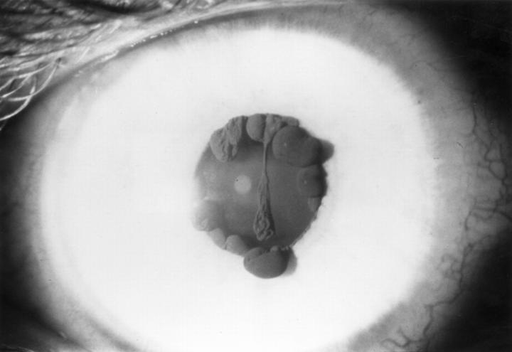
Figure 3 .
Pupillary iris pigment epithelial cyst. In this teenager, the lesions were bilateral and were noted shortly after birth. Note that some of the cysts are partly collapsed (iris flocculi).
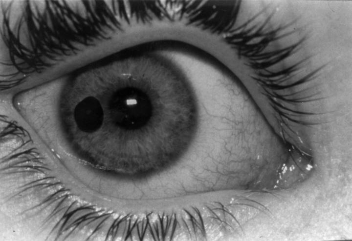
Figure 4 .
Free floating iris pigment epithelial cyst in the anterior chamber in a 12 year old girl.
Figure 5 .
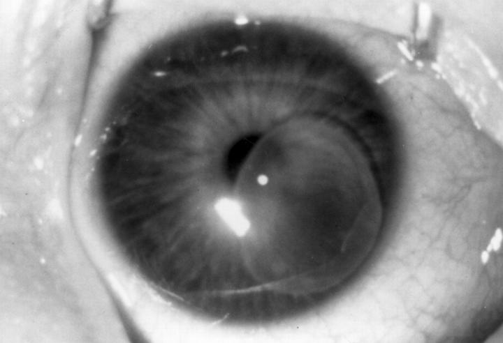
Iris stromal cyst located inferonasally in the right eye of an infant.
Figure 6 .
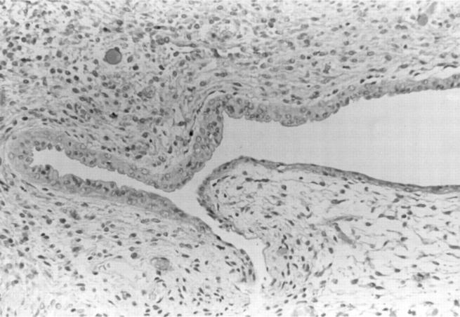
Histopathology of iris stromal cyst showing a thick wall and a lumen lined by non-keratinising epithelium similar to conjunctiva (haematoxylin and eosin, original magnification ×25).
Figure 7 .
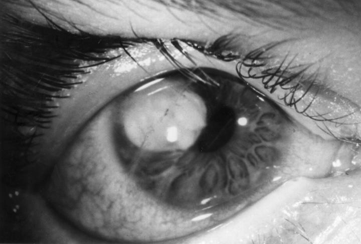
Epithelial downgrowth cyst secondary to perforating corneal trauma in an 11 year old girl. The progressively enlarging lesion was removed by a sector iridectomy.
Selected References
These references are in PubMed. This may not be the complete list of references from this article.
- Capó H., Palmer E., Nicholson D. H. Congenital cysts of the iris stroma. Am J Ophthalmol. 1993 Aug 15;116(2):228–232. doi: 10.1016/s0002-9394(14)71291-7. [DOI] [PubMed] [Google Scholar]
- GUERRY D., 3rd, WIESINGER H. Spontaneous cyst of the iris stroma. Am J Ophthalmol. 1957 Jul;44(1):106–107. doi: 10.1016/0002-9394(57)91962-1. [DOI] [PubMed] [Google Scholar]
- Grutzmacher R. D., Lindquist T. D., Chittum M. E., Bunt-Milam A. H., Kalina R. E. Congenital iris cysts. Br J Ophthalmol. 1987 Mar;71(3):227–234. doi: 10.1136/bjo.71.3.227. [DOI] [PMC free article] [PubMed] [Google Scholar]
- KLIEN B. A., TANNER G. S. Congenital epithelial cyst of the iris stroma. Am J Ophthalmol. 1963 Feb;55:291–295. doi: 10.1016/0002-9394(63)92685-0. [DOI] [PubMed] [Google Scholar]
- Lewis R. A., Merin L. M. Iris flocculi and familial aortic dissection. Arch Ophthalmol. 1995 Oct;113(10):1330–1331. doi: 10.1001/archopht.1995.01100100118041. [DOI] [PubMed] [Google Scholar]
- Lois N., Shields C. L., Shields J. A., Mercado G., De Potter P. Primary iris stromal cysts. A report of 17 cases. Ophthalmology. 1998 Jul;105(7):1317–1322. doi: 10.1016/S0161-6420(98)97041-5. [DOI] [PubMed] [Google Scholar]
- Lois N., Shields C. L., Shields J. A., Mercado G. Primary cysts of the iris pigment epithelium. Clinical features and natural course in 234 patients. Ophthalmology. 1998 Oct;105(10):1879–1885. doi: 10.1016/S0161-6420(98)91034-X. [DOI] [PubMed] [Google Scholar]
- MAKLEY T. A., Jr, KING G. L. Multiple cysts of the iris and ciliary body simulating a malignant melanoma. Trans Am Acad Ophthalmol Otolaryngol. 1958 May-Jun;62(3):441–443. [PubMed] [Google Scholar]
- Mullaney J., Fitzpatrick C. Idiopathic cyst of the iris stroma. Am J Ophthalmol. 1973 Jul;76(1):64–68. doi: 10.1016/0002-9394(73)90012-3. [DOI] [PubMed] [Google Scholar]
- Naumann G. O., Rummelt V. Block excision of cystic and diffuse epithelial ingrowth of the anterior chamber. Report on 32 consecutive patients. Arch Ophthalmol. 1992 Feb;110(2):223–227. doi: 10.1001/archopht.1992.01080140079031. [DOI] [PubMed] [Google Scholar]
- Naumann G. O., Rummelt V. Congenital nonpigmented epithelial iris cyst removed by block-excision. Graefes Arch Clin Exp Ophthalmol. 1990;228(5):392–397. doi: 10.1007/BF00927248. [DOI] [PubMed] [Google Scholar]
- Naumann G., Green W. R. Spontaneous nonpigmented iris cysts. Arch Ophthalmol. 1967 Oct;78(4):496–500. doi: 10.1001/archopht.1967.00980030498016. [DOI] [PubMed] [Google Scholar]
- Paridaens A. D., Deuble K., McCartney A. C. Spontaneous congenital non-pigmented epithelial cysts of the iris stroma. Br J Ophthalmol. 1992 Jan;76(1):39–42. doi: 10.1136/bjo.76.1.39. [DOI] [PMC free article] [PubMed] [Google Scholar]
- Roy F. H., Hanna C. Spontaneous congenital iris cyst. Am J Ophthalmol. 1971 Jul 30;72(1):97–108. doi: 10.1016/0002-9394(71)91596-0. [DOI] [PubMed] [Google Scholar]
- Rummelt V., Naumann G. O. Blockexzision mit tektonischer Korneoskleralplastik wegen zystischer und/oder diffuser Epithelinvasion des vorderen Augenabschnitts. Bericht über 51 konsekutive Patienten (1980-1996). Klin Monbl Augenheilkd. 1997 Nov;211(5):312–323. doi: 10.1055/s-2008-1035141. [DOI] [PubMed] [Google Scholar]
- Rummelt V., Rummelt C., Naumann G. O. Congenital nonpigmented epithelial iris cyst after amniocentesis. Clinicopathologic report on two children. Ophthalmology. 1993 May;100(5):776–781. doi: 10.1016/s0161-6420(93)31576-9. [DOI] [PubMed] [Google Scholar]
- Rush J. A., Weinstein G. W., Meriwether W. A. Spontaneous nonpigmented iris cyst. Arch Ophthalmol. 1982 Feb;100(2):304–305. doi: 10.1001/archopht.1982.01030030306016. [DOI] [PubMed] [Google Scholar]
- Shields C. L., Shields J. A., Cook G. R., Von Fricken M. A., Augsburger J. J. Differentiation of adenoma of the iris pigment epithelium from iris cyst and melanoma. Am J Ophthalmol. 1985 Nov 15;100(5):678–681. doi: 10.1016/0002-9394(85)90623-3. [DOI] [PubMed] [Google Scholar]
- Shields J. A., Eagle R. C., Jr, Shields C. L., De Potter P., Poliak J. G. Natural course and histopathologic findings of lacrimal gland choristoma of the iris and ciliary body. Am J Ophthalmol. 1995 Feb;119(2):219–224. doi: 10.1016/s0002-9394(14)73876-0. [DOI] [PubMed] [Google Scholar]
- Shields J. A., Eagle R. C., Jr, Shields C. L., Potter P. D. Congenital neoplasms of the nonpigmented ciliary epithelium (medulloepithelioma). Ophthalmology. 1996 Dec;103(12):1998–2006. doi: 10.1016/s0161-6420(96)30394-1. [DOI] [PubMed] [Google Scholar]
- Shields J. A., Kline M. W., Augsburger J. J. Primary iris cysts: a review of the literature and report of 62 cases. Br J Ophthalmol. 1984 Mar;68(3):152–166. doi: 10.1136/bjo.68.3.152. [DOI] [PMC free article] [PubMed] [Google Scholar]
- Shields J. A. Primary cysts of the iris. Trans Am Ophthalmol Soc. 1981;79:771–809. [PMC free article] [PubMed] [Google Scholar]
- Shields J. A., Shields C. L., DePotter P., Wagner R. S., Caputo A. R. Free-floating cyst in the anterior chamber of the eye. J Pediatr Ophthalmol Strabismus. 1996 Nov-Dec;33(6):330–331. doi: 10.3928/0191-3913-19961101-12. [DOI] [PubMed] [Google Scholar]
- Sugar H. S., Nathan L. E. Congenital epithelial cysts of the iris stroma. Ann Ophthalmol. 1982 May;14(5):483–485. [PubMed] [Google Scholar]
- Waeltermann J. M., Hettinger M. E., Cibis G. W. Congenital cysts of the iris stroma. Am J Ophthalmol. 1985 Oct 15;100(4):549–554. doi: 10.1016/0002-9394(85)90680-4. [DOI] [PubMed] [Google Scholar]
- Witschel H., Berndt K., Mackensen G. Angeborene Stromazyste der Iris. Ein klinisch-histopathologischer Fallbericht. Klin Monbl Augenheilkd. 1979 Apr;174(4):572–576. [PubMed] [Google Scholar]


