

Full Text
The Full Text of this article is available as a PDF (199.9 KB).
Figure 1 .

Fundus photograph on hospital day 4. Left eye exhibits subretinal exudative material detaching two thirds of the retina. The arrow indicates the optic disc.
Figure 2 .
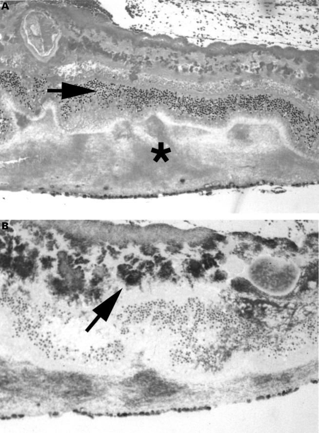
Histological sections of evisceration specimen. (A) Haematoxylin and eosin stain (×40) reveals coagulative necrosis of the inner retina. Arrow indicates outer nuclear layer. Asterisk indicates subretinal exudate with virtually no inflammatory cells. (B) Gram stain (×40) shows numerous Gram positive cocci in clusters and individually in the inner retina (arrow) and the subretinal space.