Abstract
AIMS—To map the thickness, elevation (anterior and posterior corneal surface), and axial curvature of the cornea in normal eyes with the Orbscan corneal topography system. METHODS—94 eyes of 51 normal subjects were investigated using the Orbscan corneal topography system. The anterior and posterior corneal elevation maps were classified into regular ridge, irregular ridge, incomplete ridge, island, and unclassified patterns, and the axial power maps were grouped into round, oval, symmetric bow tie, asymmetric bow tie, and irregular patterns. The pachymetry patterns were designated as round, oval, decentred round, and decentred oval. RESULTS—The thinnest point on the cornea was located at an average of 0.90 (SD 0.51) mm from visual axis and had an average thickness of 0.55 (0.03) mm. In 69.57% of eyes, this point was located in the inferotemporal quadrant, followed by the superotemporal quadrant in 23.91%, the inferonasal quadrant in 4.35%, and the superonasal quadrant in 2.17%. Among the nine regions of the cornea evaluated (central, superotemporal, temporal, inferotemporal, inferior, inferonasal, nasal, superonasal, and superior) the central cornea had the lowest average thickness (0.56 (0.03) mm) and the superior cornea had the greatest average thickness (0.64 (0.03) mm). The mean simulated keratometry (SimK) was 44.24 (1.61)/43.31 (1.66) dioptres (D) and the mean astigmatism was 0.90 (0.41) D. Island (71.74%) was the most common elevation pattern observed in the anterior corneal surface, followed by incomplete ridge (19.57%), regular ridge (4.34%), irregular ridge (2.17%), and unclassified (2.17%). Island (32.61%) was the most common topographic pattern in the posterior corneal surface, following by regular ridge (30.43%), incomplete ridge (23.91%), and irregular ridge (13.04%) patterns. Symmetric bow tie was the most common axial power pattern in the anterior cornea (39.13%), followed by oval (26.07%), asymmetric bow tie (23.91%), round (6.52%), and irregular (4.53%) patterns. In the pachymetry maps, 47.83% of eyes had an oval pattern, and round, decentred oval, and decentred round were observed in 41.30%, 8.70%, and 2.18% of eyes, respectively. CONCLUSION—The information on regional corneal thickness, corneal elevation and axial corneal curvature obtained with the Orbscan corneal topography system from normal eyes provides a reference for comparison with diseased corneas. The Orbscan corneal topography system is a useful tool to evaluate both corneal topography and corneal thickness.
Full Text
The Full Text of this article is available as a PDF (163.1 KB).
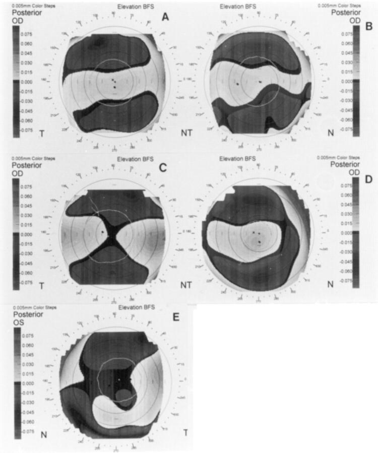
Figure 1 .
Elevation patterns of the posterior corneal surface: (A) regular ridge; (B) irregular ridge; (C) incomplete ridge; (D) island; (E) unclassified.
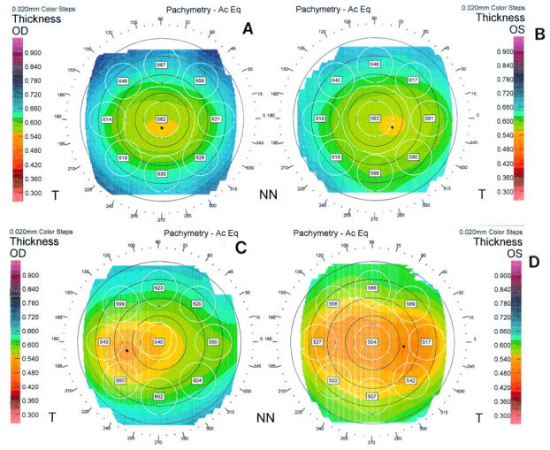
Figure 2 .

Pachymetric patterns in maps generated with the Orbscan corneal topography system: (A) oval; (B) round; (C) decentred round; (D) decentred oval.
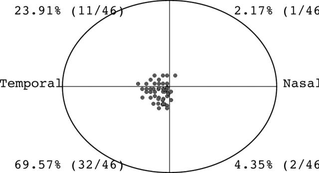
Figure 3 .
Location of the thinnest site on the cornea measured by the Orbscan corneal topography system (n=46).
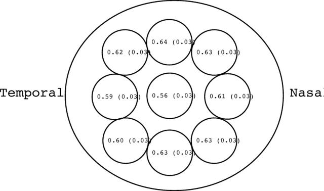
Figure 4 .
Average (SD) corneal thickness of 46 normal corneas centrally and at eight mid-peripheral locations
Selected References
These references are in PubMed. This may not be the complete list of references from this article.
- Argus W. A. Ocular hypertension and central corneal thickness. Ophthalmology. 1995 Dec;102(12):1810–1812. doi: 10.1016/s0161-6420(95)30790-7. [DOI] [PubMed] [Google Scholar]
- Auffarth G. U., Tetz M. R., Biazid Y., Völcker H. E. Measuring anterior chamber depth with Orbscan Topography System. J Cataract Refract Surg. 1997 Nov;23(9):1351–1355. doi: 10.1016/s0886-3350(97)80114-9. [DOI] [PubMed] [Google Scholar]
- Belin M. W., Cambier J. L., Nabors J. R., Ratliff C. D. PAR Corneal Topography System (PAR CTS): the clinical application of close-range photogrammetry. Optom Vis Sci. 1995 Nov;72(11):828–837. doi: 10.1097/00006324-199511000-00009. [DOI] [PubMed] [Google Scholar]
- Binder P. S. Measurement of corneal curvature after corneal transplantation and radial keratotomy using standard and automated keratometry. CLAO J. 1989 Jul-Sep;15(3):201–206. [PubMed] [Google Scholar]
- Binder P. S. Videokeratography. CLAO J. 1995 Apr;21(2):133–144. [PubMed] [Google Scholar]
- Bogan S. J., Waring G. O., 3rd, Ibrahim O., Drews C., Curtis L. Classification of normal corneal topography based on computer-assisted videokeratography. Arch Ophthalmol. 1990 Jul;108(7):945–949. doi: 10.1001/archopht.1990.01070090047037. [DOI] [PubMed] [Google Scholar]
- Cheng H., Bates A. K., Wood L., McPherson K. Positive correlation of corneal thickness and endothelial cell loss. Serial measurements after cataract surgery. Arch Ophthalmol. 1988 Jul;106(7):920–922. doi: 10.1001/archopht.1988.01060140066026. [DOI] [PubMed] [Google Scholar]
- Edmund C. Posterior corneal curvature and its influence on corneal dioptric power. Acta Ophthalmol (Copenh) 1994 Dec;72(6):715–720. doi: 10.1111/j.1755-3768.1994.tb04687.x. [DOI] [PubMed] [Google Scholar]
- Fowler C. W., Dave T. N. Review of past and present techniques of measuring corneal topography. Ophthalmic Physiol Opt. 1994 Jan;14(1):49–58. doi: 10.1111/j.1475-1313.1994.tb00556.x. [DOI] [PubMed] [Google Scholar]
- Herndon L. W., Choudhri S. A., Cox T., Damji K. F., Shields M. B., Allingham R. R. Central corneal thickness in normal, glaucomatous, and ocular hypertensive eyes. Arch Ophthalmol. 1997 Sep;115(9):1137–1141. doi: 10.1001/archopht.1997.01100160307007. [DOI] [PubMed] [Google Scholar]
- Hitzenberger C. K., Baumgartner A., Drexler W., Fercher A. F. Interferometric measurement of corneal thickness with micrometer precision. Am J Ophthalmol. 1994 Oct 15;118(4):468–476. doi: 10.1016/s0002-9394(14)75798-8. [DOI] [PubMed] [Google Scholar]
- Klyce S. D., Maurice D. M. Automatic recording of corneal thickness in vitro. Invest Ophthalmol. 1976 Jul;15(7):550–553. [PubMed] [Google Scholar]
- Lemp M. A., Dilly P. N., Boyde A. Tandem-scanning (confocal) microscopy of the full-thickness cornea. Cornea. 1985;4(4):205–209. [PubMed] [Google Scholar]
- Naufal S. C., Hess J. S., Friedlander M. H., Granet N. S. Rasterstereography-based classification of normal corneas. J Cataract Refract Surg. 1997 Mar;23(2):222–230. doi: 10.1016/s0886-3350(97)80345-8. [DOI] [PubMed] [Google Scholar]
- O'Neal M. R., Polse K. A. In vivo assessment of mechanisms controlling corneal hydration. Invest Ophthalmol Vis Sci. 1985 Jun;26(6):849–856. [PubMed] [Google Scholar]
- Petroll W. M., Roy P., Chuong C. J., Hall B., Cavanagh H. D., Jester J. V. Measurement of surgically induced corneal deformations using three-dimensional confocal microscopy. Cornea. 1996 Mar;15(2):154–164. doi: 10.1097/00003226-199603000-00008. [DOI] [PubMed] [Google Scholar]
- Prydal J. I., Artal P., Woon H., Campbell F. W. Study of human precorneal tear film thickness and structure using laser interferometry. Invest Ophthalmol Vis Sci. 1992 May;33(6):2006–2011. [PubMed] [Google Scholar]
- Remón L., Cristóbal J. A., Castillo J., Palomar T., Palomar A., Pérez J. Central and peripheral corneal thickness in full-term newborns by ultrasonic pachymetry. Invest Ophthalmol Vis Sci. 1992 Oct;33(11):3080–3083. [PubMed] [Google Scholar]
- Salz J. J., Azen S. P., Berstein J., Caroline P., Villasenor R. A., Schanzlin D. J. Evaluation and comparison of sources of variability in the measurement of corneal thickness with ultrasonic and optical pachymeters. Ophthalmic Surg. 1983 Sep;14(9):750–754. [PubMed] [Google Scholar]
- Villaseñor R. A., Santos V. R., Cox K. C., Harris D. F., 2nd, Lynn M., Waring G. O., 3rd Comparison of ultrasonic corneal thickness measurements before and during surgery in the prospective evaluation of Radial Keratotomy (PERK) Study. Ophthalmology. 1986 Mar;93(3):327–330. doi: 10.1016/s0161-6420(86)33746-1. [DOI] [PubMed] [Google Scholar]
- Waring G. O., 3rd, Bourne W. M., Edelhauser H. F., Kenyon K. R. The corneal endothelium. Normal and pathologic structure and function. Ophthalmology. 1982 Jun;89(6):531–590. [PubMed] [Google Scholar]
- Wilson S. E., Klyce S. D. Advances in the analysis of corneal topography. Surv Ophthalmol. 1991 Jan-Feb;35(4):269–277. doi: 10.1016/0039-6257(91)90047-j. [DOI] [PubMed] [Google Scholar]
- Yaylali V., Kaufman S. C., Thompson H. W. Corneal thickness measurements with the Orbscan Topography System and ultrasonic pachymetry. J Cataract Refract Surg. 1997 Nov;23(9):1345–1350. doi: 10.1016/s0886-3350(97)80113-7. [DOI] [PubMed] [Google Scholar]


