Full Text
The Full Text of this article is available as a PDF (232.2 KB).
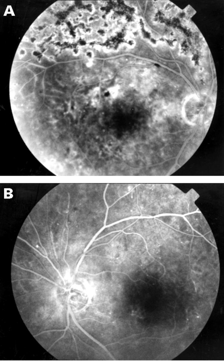
Figure 1 .

June 1996: fundus fluorescein angiogram. (A) Right eye, late venous phase. There is ischaemia of the superotemporal retina of the right eye. Superior panretinal laser scars and disc leakage from residual new vessels can be seen. (B) Left eye, mid venous phase. The left eye also shows some areas of capillary non-perfusion. There is hyperfluorescence at the superior aspect of the left disc but no characteristic leakage features of proliferative diabetic retinopathy. Note the small window defect inferonasal to the left fovea from an area of retinal pigment epithelial atrophy.
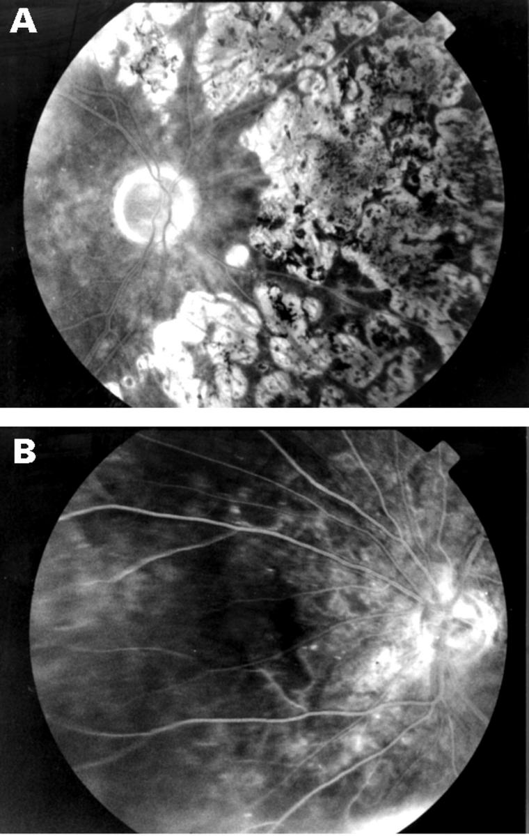
Figure 2 .

April 1998: fundus fluorescein angiogram. (A) Right eye, late phase. After multiple sessions of panretinal laser the right eye shows regression of disc new vessels with no residual leakage. (B) Left eye, mid venous phase. Following cataract surgery the hyperfluorescence at the superior aspect of the left disc is unchanged. Worsening ischaemia of the nasal retina is evident but is not accompanied by progression to proliferative diabetic retinopathy.


