Abstract
AIM—It appears from the literature that no standardised examination exists for patients with functional nasolacrimal duct obstruction. The role of dacryocystography and lacrimal scintigraphy was compared in the diagnosis and management of these patients. METHOD—Patients who were clinically diagnosed as having unilateral or bilateral functional nasolacrimal duct obstruction were prospectively entered into the study and data collected over 12 months in Moorfields Eye Hospital and Whipps Cross Hospital, London. All cases had, on separate occasions, a standardised dacryocystogram with delayed erect films and a lacrimal drainage scintigram. RESULTS—45 lacrimal systems of 32 patients (mean age 62 years; 59% male) fulfilled the inclusion criteria. Abnormalities were detected with dacryocystography in 93% of systems and with lacrimal drainage scintigraphy in 95% of systems. Based on the results of previous quantitative studies, the positive scintigrams were subdivided into those demonstrating prelacrimal sac delay (13%), delay at the lacrimal sac/duct junction (35%), or delay within the duct (47%). Combining the two imaging techniques increased the sensitivity to 98%. CONCLUSIONS—Both investigations are very sensitive at detecting abnormalities in patients with a clinical diagnosis of functional nasolacrimal duct obstruction. Lacrimal drainage scintigraphy is a slightly more sensitive test, but missed an abnormality detected by dacryocystography in two (4%) systems. A combination of the two techniques gives the highest sensitivity with maximum anatomical and physiological information but, in clinical practice, it is reasonable to perform a dacryocystogram initially and proceed to scintigraphy only if contrast radiography is normal.
Full Text
The Full Text of this article is available as a PDF (98.9 KB).
Figure 1 .
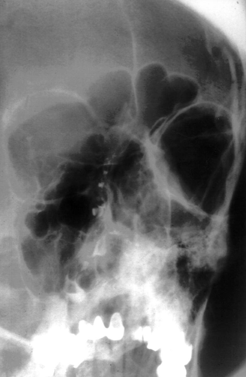
Delayed erect radiograph demonstrating retained contrast in the right lacrimal system; the left system is normal.
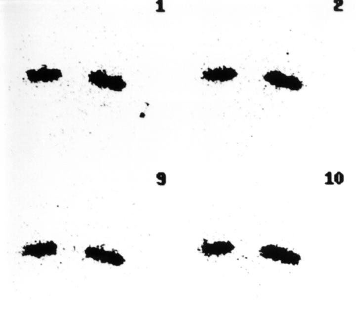
Figure 2 .
Normal lacrimating scintigram with rapid clearance of tracer in dynamic study (pictures at 10, 20, 90, and 100 seconds).
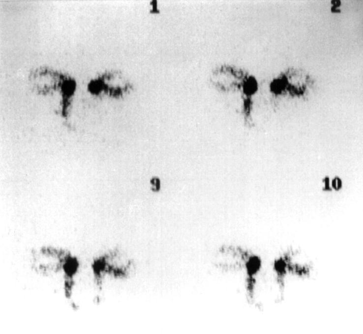
Figure 3 .
Dynamic study showing system with "presac delay".
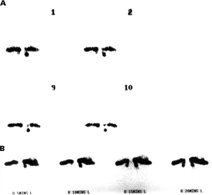
Figure 4 .
(A) Dynamic study with rapid entry of tracer into the lacrimal sac on the right and upper nasolacrimal duct on the left. (B) Static films (same patient as (A)) confirming "preductal delay" on the right and "intraduct delay" on the left.
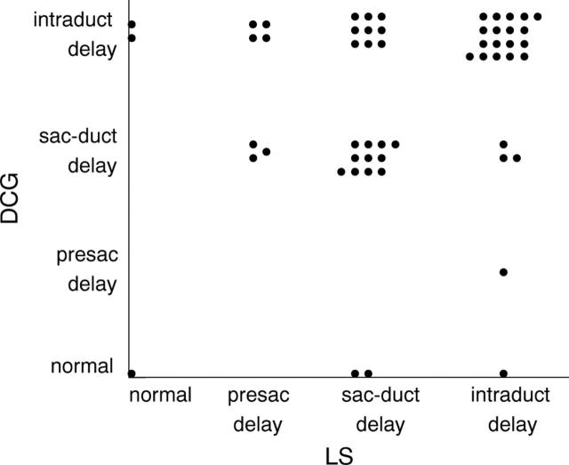
Figure 5 .
Graphic representation of main site of delay in dacryocystography and lacrimal scintigraphy.
Selected References
These references are in PubMed. This may not be the complete list of references from this article.
- Amanat L. A., Hilditch T. E., Kwok C. S. Lacrimal scintigraphy. II. Its role in the diagnosis of epiphora. Br J Ophthalmol. 1983 Nov;67(11):720–728. doi: 10.1136/bjo.67.11.720. [DOI] [PMC free article] [PubMed] [Google Scholar]
- Brown M., El Gammal T. A., Luxenberg M. N., Eubig C. The value, limitations, and applications of nuclear dacryocystography. Semin Nucl Med. 1981 Oct;11(4):250–257. doi: 10.1016/s0001-2998(81)80022-0. [DOI] [PubMed] [Google Scholar]
- Carlton W. H., Trueblood J. H., Rossomondo R. M. Clinical evaluation of microscintigraphy of the lacrimal drainage apparatus. J Nucl Med. 1973 Feb;14(2):89–92. [PubMed] [Google Scholar]
- Chaudhuri R. K., Saparoff G. R., Dolan K. D., Chaudhuri T. K. A comparative study of contrast dacryocystogram and nuclear dacryocystogram. J Nucl Med. 1975 Jul;16(7):605–608. [PubMed] [Google Scholar]
- Chavis R. M., Welham R. A., Maisey M. N. Quantitative lacrimal scintillography. Arch Ophthalmol. 1978 Nov;96(11):2066–2068. doi: 10.1001/archopht.1978.03910060454013. [DOI] [PubMed] [Google Scholar]
- Conway S. T. Evaluation and management of "functional" nasolacrimal blockage: results of a survey of the American Society of Ophthalmic Plastic and Reconstructive surgery. Ophthal Plast Reconstr Surg. 1994 Sep;10(3):185–188. [PubMed] [Google Scholar]
- Hurwitz J. J., Maisey M. N., Welham R. A. Quantitative lacrimal scintillography. I. Method and physiological application. Br J Ophthalmol. 1975 Jun;59(6):308–312. doi: 10.1136/bjo.59.6.308. [DOI] [PMC free article] [PubMed] [Google Scholar]
- Hurwitz J. J., Welham R. A., Lloyd G. A. The role of intubation macro-dacryocystography in management of problems of the lacrimal system. Can J Ophthalmol. 1975 Jul;10(3):361–366. [PubMed] [Google Scholar]
- Lloyd G. A., Jones B. R., Welham R. A. Intubation macrodacryocystography. Br J Ophthalmol. 1972 Aug;56(8):600–603. doi: 10.1136/bjo.56.8.600. [DOI] [PMC free article] [PubMed] [Google Scholar]
- Rose J. D., Clayton C. B. Scintigraphy and contrast radiography for epiphora. Br J Radiol. 1985 Dec;58(696):1183–1186. doi: 10.1259/0007-1285-58-696-1183. [DOI] [PubMed] [Google Scholar]
- Rossomondo R. M., Carlton W. H., Trueblood J. H., Thomas R. P. A new method of evaluating lacrimal drainage. Arch Ophthalmol. 1972 Nov;88(5):523–525. doi: 10.1001/archopht.1972.01000030525010. [DOI] [PubMed] [Google Scholar]
- Saraç K., Hepşen I. F., Bayramlar H., Uguralp M., Toksöz M., Baysal T. Computed tomography dacryocystography. Eur J Radiol. 1995 Jan;19(2):128–131. doi: 10.1016/0720-048x(94)00385-p. [DOI] [PubMed] [Google Scholar]


