Full Text
The Full Text of this article is available as a PDF (244.4 KB).
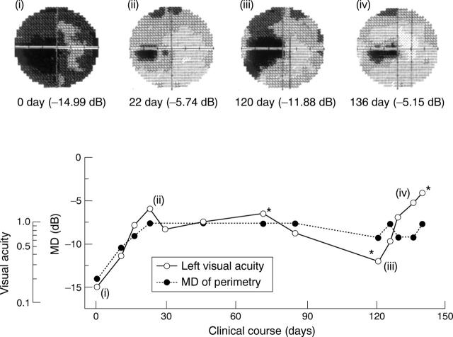
Figure 1 .
(Top) Raw images of Humphrey 30-2 visual fields in the left eye. (Bottom) The relation between clinical course and mean deviation (MD) of Humphrey 30-2 visual field and visual acuity in the left eye. Asterisks indicate the day in which multifocal ERG was analysed. The roman numerals correspond with raw images in the upper part of the figure.
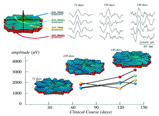
Figure 2 .
(Top left) In the multifocal ERG, the fundus was divided into four foci. (Top right) Sum of the amplitudes in each foci was altered during the clinical course. (Bottom) The three dimensional topography and sum of the amplitudes in each of four foci of the m-ERG were indicated.


