Abstract
AIM—To assess serial, simultaneous stereo optic disc images by four methods for glaucomatous progression. METHODS—Using varying techniques, two ophthalmologists assessed serial optic disc images of 52 eyes from 27 patients with a mean duration between images of 18 months. The neuroretinal rim width was qualitatively assessed by four assessment methods and compared with quantitative rim measurements made using PC based software. RESULTS—The highest sensitivity of 83% was achieved using computerised stereo chronoscopy. CONCLUSION—Stereo chronoscopy improved the detection of subtle optic disc changes when compared with simpler assessment techniques.
Full Text
The Full Text of this article is available as a PDF (78.0 KB).
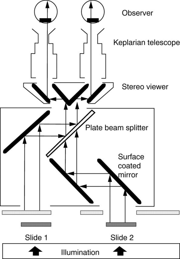
Figure 1 .

Schematic diagram of the optical/mechanical stereo flicker chronoscopy comparator.
Selected References
These references are in PubMed. This may not be the complete list of references from this article.
- Bengtsson B., Krakau C. E. Flicker comparison of fundus photographs. A technical note. Acta Ophthalmol (Copenh) 1979 Jun;57(3):503–506. doi: 10.1111/j.1755-3768.1979.tb01834.x. [DOI] [PubMed] [Google Scholar]
- Goldmann H., Lotmar W. Quantitative studies in stereochronoscopy (Sc): application to the disc in glaucoma. I. Phenomenology. Graefes Arch Clin Exp Ophthalmol. 1984;222(1):38–42. doi: 10.1007/BF02133776. [DOI] [PubMed] [Google Scholar]
- Heijl A., Bengtsson B. Diagnosis of early glaucoma with flicker comparisons of serial disc photographs. Invest Ophthalmol Vis Sci. 1989 Nov;30(11):2376–2384. [PubMed] [Google Scholar]
- Takamoto T., Schwartz B. Stereochronometry: quantitative measurement of optic disc cup changes. Invest Ophthalmol Vis Sci. 1985 Oct;26(10):1445–1449. [PubMed] [Google Scholar]


