Abstract
AIMS—To investigate the frequency of corneal guttata in patients with a corneal dystrophy resulting from an Arg124His (R124H) mutation of βig-h3 gene. METHODS—Slit lamp examination was performed on 30 eyes with corneal dystrophy from a genetically confirmed βig-h3 R124H mutation and on 50 age matched control eyes. The stage of the corneal dystrophy was classified as stage 0, I, or II and the degree of guttata was classified as none, mild, or severe. Specular microscopic examinations were performed to evaluate the morphology of the corneal endothelium. RESULTS—Slit lamp examination disclosed the presence of corneal guttata in 21 eyes (70%) of the 30 eyes with the corneal dystrophy, but in only one (2%) of the 50 eyes in the age matched control group (p<0.001, χ2 with Yates's correction). Of the 12 eyes with stage I βig-h3 R124H corneal dystrophy, seven had no corneal guttata and five had a mild degree of guttata. Of the 18 eyes with stage II, the degree of guttata was none in two, mild in nine, and severe in seven. The degree of corneal guttata was significantly related to the stage of the corneal dystrophy (p<0.0001, Kruskul-Wallis test ANOVA on ranks). There was no significant differences between eyes with βig-h3 R124H corneal dystrophy and normal eyes in cell density, coefficient of variation, and cell hexagonality of corneal endothelium. CONCLUSION—Corneal guttata are one of the characteristics of the corneal dystrophy resulting from βig-h3 R124H mutation.
Full Text
The Full Text of this article is available as a PDF (183.9 KB).
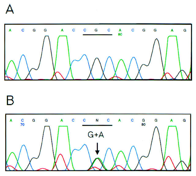
Figure 1 .
Molecular analysis of patients with corneal dystrophy from a heterozygous R124H mutation of βig-h3 gene. The sequence reveals unaffected patients (A) and affected patients (B). G to A transition (CGC to CAC) was found at position 418 in the affected patients.
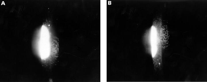
Figure 2 .
Staging of corneal dystrophy. (A) Stage I of the dystrophy is characterised by isolated deposits in an otherwise clear cornea. (B) Stage II is characterised by moderate to abundant deposits and corneal haze.
Figure 3 .
Grading of corneal guttata. (A) Mild corneal guttata are characterised by a small number of guttata formations. (B) Severe corneal guttata are characterised by abundant guttata formations.
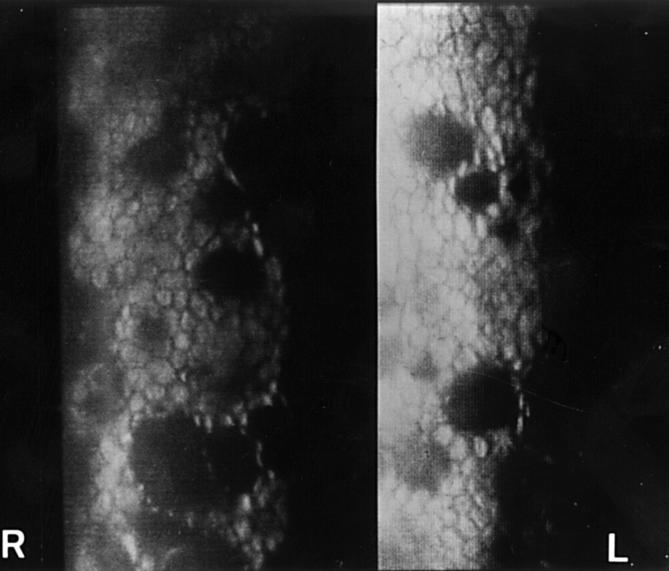
Figure 4 .
Specular microscopic photograph. Severe corneal guttata were observed in the right eye (R) and mild guttata in the left eye (L) of the same 64 year old patient with βig-h3 R124H mutation.
Selected References
These references are in PubMed. This may not be the complete list of references from this article.
- Folberg R., Alfonso E., Croxatto J. O., Driezen N. G., Panjwani N., Laibson P. R., Boruchoff S. A., Baum J., Malbran E. S., Fernandez-Meijide R. Clinically atypical granular corneal dystrophy with pathologic features of lattice-like amyloid deposits. A study of these families. Ophthalmology. 1988 Jan;95(1):46–51. doi: 10.1016/s0161-6420(88)33226-4. [DOI] [PubMed] [Google Scholar]
- Hanna K. D., Pouliquen Y., Waring G. O., 3rd, Savoldelli M., Cotter J., Morton K., Menasche M. Corneal stromal wound healing in rabbits after 193-nm excimer laser surface ablation. Arch Ophthalmol. 1989 Jun;107(6):895–901. doi: 10.1001/archopht.1989.01070010917041. [DOI] [PubMed] [Google Scholar]
- Hogan M. J., Wood I., Fine M. Fuchs' endothelial dystrophy of the cornea. 29th Sanford Gifford Memorial lecture. Am J Ophthalmol. 1974 Sep;78(3):363–383. doi: 10.1016/0002-9394(74)90224-4. [DOI] [PubMed] [Google Scholar]
- Holland E. J., Daya S. M., Stone E. M., Folberg R., Dobler A. A., Cameron J. D., Doughman D. J. Avellino corneal dystrophy. Clinical manifestations and natural history. Ophthalmology. 1992 Oct;99(10):1564–1568. doi: 10.1016/s0161-6420(92)31766-x. [DOI] [PubMed] [Google Scholar]
- Konishi M., Mashima Y., Nakamura Y., Yamada M., Sugiura H. Granular-lattice (Avellino) corneal dystrophy in Japanese patients. Cornea. 1997 Nov;16(6):635–638. [PubMed] [Google Scholar]
- Munier F. L., Korvatska E., Djemaï A., Le Paslier D., Zografos L., Pescia G., Schorderet D. F. Kerato-epithelin mutations in four 5q31-linked corneal dystrophies. Nat Genet. 1997 Mar;15(3):247–251. doi: 10.1038/ng0397-247. [DOI] [PubMed] [Google Scholar]
- Okada M., Yamamoto S., Inoue Y., Watanabe H., Maeda N., Shimomura Y., Ishii Y., Tano Y. Severe corneal dystrophy phenotype caused by homozygous R124H keratoepithelin mutations. Invest Ophthalmol Vis Sci. 1998 Sep;39(10):1947–1953. [PubMed] [Google Scholar]
- Rosenwasser G. O., Sucheski B. M., Rosa N., Pastena B., Sebastiani A., Sassani J. W., Perry H. D. Phenotypic variation in combined granular-lattice (Avellino) corneal dystrophy. Arch Ophthalmol. 1993 Nov;111(11):1546–1552. doi: 10.1001/archopht.1993.01090110112035. [DOI] [PubMed] [Google Scholar]


