Abstract
BACKGROUND/AIMS—Morphological variability of the trabecular meshwork could be of considerable importance for the proper intraoperative outcome of non-perforating antiglaucomatous surgery, such as deep sclerectomy and viscocanalostomy. The aim of this study was therefore to assess qualitative and quantitative characteristics of the trabecular meshwork in glaucoma patients undergoing trabeculectomy. METHODS—Trabeculectomy specimens from 177 glaucoma patients were prepared for light microscopy; 100 specimens were found to be suitable for qualitative assessment and quantitative computerised image analysis; measurements were taken of the meridional diameter of Schlemm's canal as well as the thickness of the trabecular meshwork at different positions. RESULTS—The mean meridional diameter of Schlemm's canal was 290 µm with the smallest values in the young patients with infantile and secondary glaucomas. the thickness of the trabecular meshwork ranged between 50-70 µm in the anterior region and between 100-130 µm for the posterior portion. The thickness of the anterior meshwork significantly decreased with age. The pigmentation of excised trabecular meshwork was found to be weak or even lacking in 68 patients. In 20 glaucoma patients the uveal meshwork was covered by an endothelial layer. CONCLUSIONS—From the morphological point of view the risk of inadvertent perforation during deep sclerectomy in older, white glaucoma patients should be taken into account even by an experienced surgeon, because the anterior meshwork in these cases is very thin and trabecular pigmentation that can be used as a topographic landmark is often lacking. The functional success of non-perforating glaucoma surgery in many patients may be limited by endothelial covering of the trabecular meshwork.
Full Text
The Full Text of this article is available as a PDF (183.0 KB).
Figure 1 .
Trabecular meshwork in a 67 year old patient with primary open angle glaucoma revealing no pigmentation. The broken line shows the meridional diameter of Schlemm's canal (SC), lines 1-4 indicate the positions where the thickness of the trabecular meshwork was measured. Original magnification ×134.
Figure 2 .
Linear regression line with 95% CI describing the relation between age and thickness of the trabecular meshwork at position 1: slope is −0.44 (0.11); r2=0.16; p<0.0001.
Figure 3 .
Linear regression line with 95% CI describing the relation between age and thickness of the trabecular meshwork at position 2: slope is −0.42 (0.15); r2=0.07; p=0.006.
Figure 4 .
Linear regression line with 95% CI describing the relation between age and thickness of the trabecular meshwork at position 3: slope is −0.07 (0.23); r2=0.001; p=0.75.
Figure 5 .
Linear regression line with 95% CI describing the relation between age and meridional diameter of Schlemm's canal: slope is 1.3 (0.44); r2=0.09; p=0.004.
Figure 6 .
Trabecular meshwork in a 57 year old patient with irido-corneo-endothelial (ICE) syndrome and secondary glaucoma. The lumen of Schlemm's canal (SC) is bridged by several tissue bands. The uveal meshwork is completely covered by a thick endothelial layer (arrowheads). Original magnification ×309.
Figure 7 .
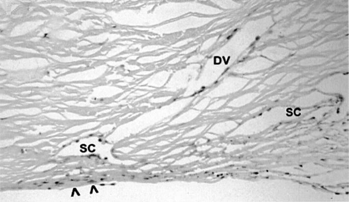
Trabecular meshwork in a 72 year old patient with primary open angle glaucoma and history of laser trabeculoplasty. Schlemm's canal (SC) consists of separated lumina; a radially running drainage vessel (DV) meets the anterior part of Schlemm's canal. The anterior meshwork shows a loss of intertrabecular space and an endothelial covering (arrows). Original magnification ×244.
Figure 8 .
Trabecular meshwork in a 65 year old patient with exfoliative glaucoma showing moderate pigmentation. A "bridge-like channel" (CH) goes tangentially along with Schlemm's canal (SC) proved by serial sections. Original magnification x 259.
Selected References
These references are in PubMed. This may not be the complete list of references from this article.
- Ainsworth J. R., Lee W. R. Effects of age and rapid high-pressure fixation on the morphology of Schlemm's canal. Invest Ophthalmol Vis Sci. 1990 Apr;31(4):745–750. [PubMed] [Google Scholar]
- Alexander R. A., Grierson I., Church W. H. The effect of argon laser trabeculoplasty upon the normal human trabecular meshwork. Graefes Arch Clin Exp Ophthalmol. 1989;227(1):72–77. doi: 10.1007/BF02169830. [DOI] [PubMed] [Google Scholar]
- Allingham R. R., de Kater A. W., Ethier C. R. Schlemm's canal and primary open angle glaucoma: correlation between Schlemm's canal dimensions and outflow facility. Exp Eye Res. 1996 Jan;62(1):101–109. doi: 10.1006/exer.1996.0012. [DOI] [PubMed] [Google Scholar]
- Buller C., Johnson D. Segmental variability of the trabecular meshwork in normal and glaucomatous eyes. Invest Ophthalmol Vis Sci. 1994 Oct;35(11):3841–3851. [PubMed] [Google Scholar]
- Carassa R. G., Bettin P., Fiori M., Brancato R. Viscocanalostomy: a pilot study. Eur J Ophthalmol. 1998 Apr-Jun;8(2):57–61. doi: 10.1177/112067219800800201. [DOI] [PubMed] [Google Scholar]
- Chiou A. G., Mermoud A., Underdahl J. P., Schnyder C. C. An ultrasound biomicroscopic study of eyes after deep sclerectomy with collagen implant. Ophthalmology. 1998 Apr;105(4):746–750. doi: 10.1016/S0161-6420(98)94033-7. [DOI] [PubMed] [Google Scholar]
- Gottanka J., Johnson D. H., Martus P., Lütjen-Drecoll E. Severity of optic nerve damage in eyes with POAG is correlated with changes in the trabecular meshwork. J Glaucoma. 1997 Apr;6(2):123–132. [PubMed] [Google Scholar]
- Hamanaka T., Bill A., Ichinohasama R., Ishida T. Aspects of the development of Schlemm's canal. Exp Eye Res. 1992 Sep;55(3):479–488. doi: 10.1016/0014-4835(92)90121-8. [DOI] [PubMed] [Google Scholar]
- Khaw P. T., Siriwardena D. "New" surgical treatments for glaucoma. Br J Ophthalmol. 1999 Jan;83(1):1–2. doi: 10.1136/bjo.83.1.1. [DOI] [PMC free article] [PubMed] [Google Scholar]
- Krieglstein G. K. How new is new, and is it better? J Glaucoma. 1999 Oct;8(5):279–280. [PubMed] [Google Scholar]
- Lee W. R. Doyne Lecture. The pathology of the outflow system in primary and secondary glaucoma. Eye (Lond) 1995;9(Pt 1):1–23. doi: 10.1038/eye.1995.2. [DOI] [PubMed] [Google Scholar]
- Lütjen E., Rohen J. W. Histometrische Untersuchungen über die Kammerwinkelregion des menschlichen Auges bei verschiedenen Alterstufen und Glaukomformen. Albrecht Von Graefes Arch Klin Exp Ophthalmol. 1968;176(1):1–12. doi: 10.1007/BF00430625. [DOI] [PubMed] [Google Scholar]
- McMenamin P. G., Lee W. R., Aitken D. A. Age-related changes in the human outflow apparatus. Ophthalmology. 1986 Feb;93(2):194–209. doi: 10.1016/s0161-6420(86)33762-x. [DOI] [PubMed] [Google Scholar]
- Mermoud A., Schnyder C. C., Sickenberg M., Chiou A. G., Hédiguer S. E., Faggioni R. Comparison of deep sclerectomy with collagen implant and trabeculectomy in open-angle glaucoma. J Cataract Refract Surg. 1999 Mar;25(3):323–331. doi: 10.1016/s0886-3350(99)80079-0. [DOI] [PubMed] [Google Scholar]
- Miyazaki M., Segawa K., Urakawa Y. Age-related changes in the trabecular meshwork of the normal human eye. Jpn J Ophthalmol. 1987;31(4):558–569. [PubMed] [Google Scholar]
- Nesterov A. P., Batmanov Y. E. Study on morphology and function of the drainage area of the eye of man. Acta Ophthalmol (Copenh) 1972;50(3):337–350. doi: 10.1111/j.1755-3768.1972.tb05956.x. [DOI] [PubMed] [Google Scholar]
- Nesterov A. P., Hasanova N. H., Batmanov Y. E. Schlemm's canal and scleral spur in normal and glaucomatous eyes. Acta Ophthalmol (Copenh) 1974;52(5):634–646. doi: 10.1111/j.1755-3768.1974.tb01099.x. [DOI] [PubMed] [Google Scholar]
- Rodrigues M. M., Spaeth G. L., Donohoo P. Electron microscopy of argon laser therapy in phakic open-angle glaucoma. Ophthalmology. 1982 Mar;89(3):198–210. doi: 10.1016/s0161-6420(82)34806-x. [DOI] [PubMed] [Google Scholar]
- Rodrigues M. M., Spaeth G. L., Sivalingam E., Weinreb S. Histopathology of 150 trabeculectomy specimens in glaucoma. Trans Ophthalmol Soc U K. 1976 Jul;96(2):245–255. [PubMed] [Google Scholar]
- Rodrigues M. M., Spaeth G. L., Sivalingam E., Weinreb S. Value of trabeculectomy specimens in glaucoma. Ophthalmic Surg. 1978 Apr;9(2):29–38. [PubMed] [Google Scholar]
- Rohen J. W. New studies on the functional morphology of the trabecular meshwork and the outflow channels. Trans Ophthalmol Soc U K. 1970;89:431–447. [PubMed] [Google Scholar]
- Sanchez E., Schnyder C. C., Mermoud A. Résultats comparatifs de la sclérectomie profonde transformée en trabéculectomie et de la trabéculectomie classique. Klin Monbl Augenheilkd. 1997 May;210(5):261–264. doi: 10.1055/s-2008-1035050. [DOI] [PubMed] [Google Scholar]
- Stegmann R., Pienaar A., Miller D. Viscocanalostomy for open-angle glaucoma in black African patients. J Cataract Refract Surg. 1999 Mar;25(3):316–322. doi: 10.1016/s0886-3350(99)80078-9. [DOI] [PubMed] [Google Scholar]
- Watson P. G., Jakeman C., Ozturk M., Barnett M. F., Barnett F., Khaw K. T. The complications of trabeculectomy (a 20-year follow-up). Eye (Lond) 1990;4(Pt 3):425–438. doi: 10.1038/eye.1990.54. [DOI] [PubMed] [Google Scholar]
- Welsh N. H., DeLange J., Wasserman P., Ziémba S. L. The "deroofing" of Schlemm's canal in patients with open-angle glaucoma through placement of a collagen drainage device. Ophthalmic Surg Lasers. 1998 Mar;29(3):216–226. [PubMed] [Google Scholar]
- Zimmerman T. J., Kooner K. S., Ford V. J., Olander K. W., Mandlekorn R. M., Rawlings E. F., Leader B. J., Koskan A. J. Trabeculectomy vs. nonpenetrating trabeculectomy: a retrospective study of two procedures in phakic patients with glaucoma. Ophthalmic Surg. 1984 Sep;15(9):734–740. [PubMed] [Google Scholar]


