

Abstract
AIMS—To analyse the histopathology of classic and occult choroidal neovascular membrane surgical specimens in age related macular degeneration. METHODS—35 membranes, from a consecutive series of surgically removed choroidal neovascular membranes in age related macular degeneration, were classified as classic or occult following the guidelines of the Macular Photocoagulation Study. Membranes with classic as well as occult components were considered as mixed membranes. The membranes were serially sectioned and stained with haematoxylin and eosin, Masson trichrome, periodic acid-Schiff, and phosphotungstic acid haematoxylin stain. The correlation has been made in a masked fashion. RESULTS—31 membranes (19 classic, 10 occult, and two mixed membranes) could be analysed histologically. 18 classic choroidal neovascular membranes had a major subretinal fibrovascular component and 10 of these had an additional, minor fibrovascular component under the retinal pigment epithelium. The 10 occult membranes contained a fibrovascular component under the retinal pigment epithelium and the two mixed membranes contained fibrovascular tissue on both sides of the retinal pigment epithelium. Fibrin and remains of outer segments tended to occur at the lateral edges of classic membranes and to cover the inner surface of occult membranes. CONCLUSION—Classic choroidal neovascularisation in age related macular degeneration is predominantly composed of subretinal fibrovascular tissue while occult choroidal neovascularisation is composed of fibrovascular tissue at the choroidal side of the retinal pigment epithelium.
Full Text
The Full Text of this article is available as a PDF (191.3 KB).
Figure 1 .

(A) Early phase and (B) late phase fluorescein angiogram of a classic choroidal neovascular membrane in age related macular degeneration from a 83 year old patient. (C) Early phase and (D) late phase fluorescein angiogram of an occult choroidal neovascular membrane in age related macular degeneration from a 75 year old patient.
Figure 2 .
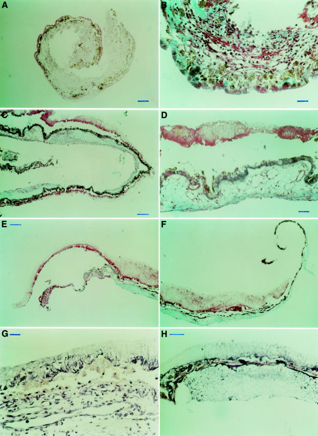
(A) PAS stain of a classic membrane, corresponding to Figure 1A and B, indicating a major subretinal and a minor sub-RPE fibrovascular component. Both these components are separated by diffuse drusen that stained as a PAS positive line. Bar = 100 µm. (B) MTC stain, detail of (A). Bar = 25 µm. (C) MTC stain of an occult choroidal neovascular membrane, corresponding to Figure 1C and D, shows a sub-RPE fibrovascular membrane and remains of outer segments covering the inner surface of the membrane. Bar = 100 µm. (D) MTC stain, detail of (C). Bar = 25 µm. (E, F) MTC stain of another occult choroidal neovascular membrane; fibrovascular tissue is seen at the choroidal side of the retinal pigment epithelium and remains of outer segment material cover the inner surface of the membrane. Bar = 100 µm. (G) PTAH stain of an occult choroidal neovascular membrane. Fibrin is seen interspersed with remains of outer segments. Bar = 25 µm. (H) PTAH stain of an occult choroidal neovascular membrane. A serofibrinous pigment epithelial detachment is seen. Bar = 50 µm.
Selected References
These references are in PubMed. This may not be the complete list of references from this article.
- Bressler N. M., Bressler S. B., Fine S. L. Age-related macular degeneration. Surv Ophthalmol. 1988 May-Jun;32(6):375–413. doi: 10.1016/0039-6257(88)90052-5. [DOI] [PubMed] [Google Scholar]
- Bressler N. M., Bressler S. B., Gragoudas E. S. Clinical characteristics of choroidal neovascular membranes. Arch Ophthalmol. 1987 Feb;105(2):209–213. doi: 10.1001/archopht.1987.01060020063030. [DOI] [PubMed] [Google Scholar]
- Bressler S. B., Silva J. C., Bressler N. M., Alexander J., Green W. R. Clinicopathologic correlation of occult choroidal neovascularization in age-related macular degeneration. Arch Ophthalmol. 1992 Jun;110(6):827–832. doi: 10.1001/archopht.1992.01080180099035. [DOI] [PubMed] [Google Scholar]
- Chang T. S., Freund K. B., de la Cruz Z., Yannuzzi L. A., Green W. R. Clinicopathologic correlation of choroidal neovascularization demonstrated by indocyanine green angiography in a patient with retention of good vision for almost four years. Retina. 1994;14(2):114–124. doi: 10.1097/00006982-199414020-00004. [DOI] [PubMed] [Google Scholar]
- Das A., Puklin J. E., Frank R. N., Zhang N. L. Ultrastructural immunocytochemistry of subretinal neovascular membranes in age-related macular degeneration. Ophthalmology. 1992 Sep;99(9):1368–1376. doi: 10.1016/s0161-6420(92)31792-0. [DOI] [PubMed] [Google Scholar]
- Gass J. D. Biomicroscopic and histopathologic considerations regarding the feasibility of surgical excision of subfoveal neovascular membranes. Am J Ophthalmol. 1994 Sep 15;118(3):285–298. [PubMed] [Google Scholar]
- Green W. R., Enger C. Age-related macular degeneration histopathologic studies. The 1992 Lorenz E. Zimmerman Lecture. Ophthalmology. 1993 Oct;100(10):1519–1535. doi: 10.1016/s0161-6420(93)31466-1. [DOI] [PubMed] [Google Scholar]
- Green W. R., McDonnell P. J., Yeo J. H. Pathologic features of senile macular degeneration. Ophthalmology. 1985 May;92(5):615–627. [PubMed] [Google Scholar]
- Grossniklaus H. E., Gass J. D. Clinicopathologic correlations of surgically excised type 1 and type 2 submacular choroidal neovascular membranes. Am J Ophthalmol. 1998 Jul;126(1):59–69. doi: 10.1016/s0002-9394(98)00145-7. [DOI] [PubMed] [Google Scholar]
- Grossniklaus H. E., Green W. R. Histopathologic and ultrastructural findings of surgically excised choroidal neovascularization. Submacular Surgery Trials Research Group. Arch Ophthalmol. 1998 Jun;116(6):745–749. doi: 10.1001/archopht.116.6.745. [DOI] [PubMed] [Google Scholar]
- Grossniklaus H. E., Hutchinson A. K., Capone A., Jr, Woolfson J., Lambert H. M. Clinicopathologic features of surgically excised choroidal neovascular membranes. Ophthalmology. 1994 Jun;101(6):1099–1111. doi: 10.1016/s0161-6420(13)31216-0. [DOI] [PubMed] [Google Scholar]
- Grossniklaus H. E., Martinez J. A., Brown V. B., Lambert H. M., Sternberg P., Jr, Capone A., Jr, Aaberg T. M., Lopez P. F. Immunohistochemical and histochemical properties of surgically excised subretinal neovascular membranes in age-related macular degeneration. Am J Ophthalmol. 1992 Oct 15;114(4):464–472. doi: 10.1016/s0002-9394(14)71859-8. [DOI] [PubMed] [Google Scholar]
- Lopez P. F., Grossniklaus H. E., Lambert H. M., Aaberg T. M., Capone A., Jr, Sternberg P., Jr, L'Hernault N. Pathologic features of surgically excised subretinal neovascular membranes in age-related macular degeneration. Am J Ophthalmol. 1991 Dec 15;112(6):647–656. doi: 10.1016/s0002-9394(14)77270-8. [DOI] [PubMed] [Google Scholar]
- Lopez P. F., Lambert H. M., Grossniklaus H. E., Sternberg P., Jr Well-defined subfoveal choroidal neovascular membranes in age-related macular degeneration. Ophthalmology. 1993 Mar;100(3):415–422. doi: 10.1016/s0161-6420(93)31657-x. [DOI] [PubMed] [Google Scholar]
- Sarks J. P., Sarks S. H., Killingsworth M. C. Morphology of early choroidal neovascularisation in age-related macular degeneration: correlation with activity. Eye (Lond) 1997;11(Pt 4):515–522. doi: 10.1038/eye.1997.137. [DOI] [PubMed] [Google Scholar]
- Sarks S. H. New vessel formation beneath the retinal pigment epithelium in senile eyes. Br J Ophthalmol. 1973 Dec;57(12):951–965. doi: 10.1136/bjo.57.12.951. [DOI] [PMC free article] [PubMed] [Google Scholar]
- Small M. L., Green W. R., Alpar J. J., Drewry R. E. Senile mecular degeneration. A clinicopathologic correlation of two cases with neovascularization beneath the retinal pigment epithelium. Arch Ophthalmol. 1976 Apr;94(4):601–607. doi: 10.1001/archopht.1976.03910030287008. [DOI] [PubMed] [Google Scholar]
- de Juan E., Jr, Machemer R. Vitreous surgery for hemorrhagic and fibrous complications of age-related macular degeneration. Am J Ophthalmol. 1988 Jan 15;105(1):25–29. doi: 10.1016/0002-9394(88)90116-x. [DOI] [PubMed] [Google Scholar]