Abstract
BACKGROUND/AIMS—"Hurricane keratopathy" is the name given to the whorl pattern, highlighted with fluorescein, seen in situations where corneal epithelial cell turnover is exaggerated. Although the condition is well described, follow up data on patients with this condition and its sequelae have only been reported in corneal graft patients. The aim was to study the clinical course of hurricane keratopathy in corneal graft patients and contact lens wearers, and to document any sequelae of this condition. METHODS—Hurricane keratopathy, occurring in 20 eyes with corneal grafts and 16 eyes (six bilateral) wearing rigid gas permeable contact lenses, was studied and followed. The occurrence, pattern, progress, resolution, and residual effects of the whorls were noted. RESULTS—Hurricane keratopathy was noted to occur in grafts as previously reported and also in contact lens wearers, which has hitherto not been reported. The whorls usually appeared within the first 3 weeks postoperatively and persisted up to 4 months. A small epithelial defect (11.1%), heaped epithelial cells (5.6%), and a nebular grade opacity (2.8%), were the only significant sequelae noted at the epicentre of the whorls. Resolution occurred from the periphery towards the centre of the cornea. CONCLUSIONS—The whorl pattern is sustained as long as the stimulus for increased cell turnover is maintained. Once this stimulus is eliminated, the pattern tends to resolve spontaneously.
Full Text
The Full Text of this article is available as a PDF (120.8 KB).
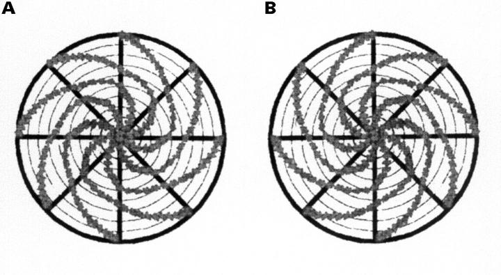
Figure 1 .
(A) Diagram representing a clockwise whorl. When a whorl on either eye conformed to the appearance in (A), it was deemed to be a clockwise whorl. (B) Diagram representing an anti-clockwise whorl. When a whorl on either eye conformed to the appearance in (B), it was deemed to be an anti-clockwise whorl.
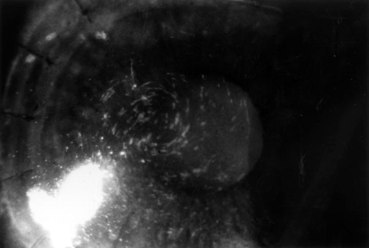
Figure 2 .
A temporally located clockwise whorl in the right eye of a patient with a corneal graft. There is no epithelial defect in the centre of the whorl.
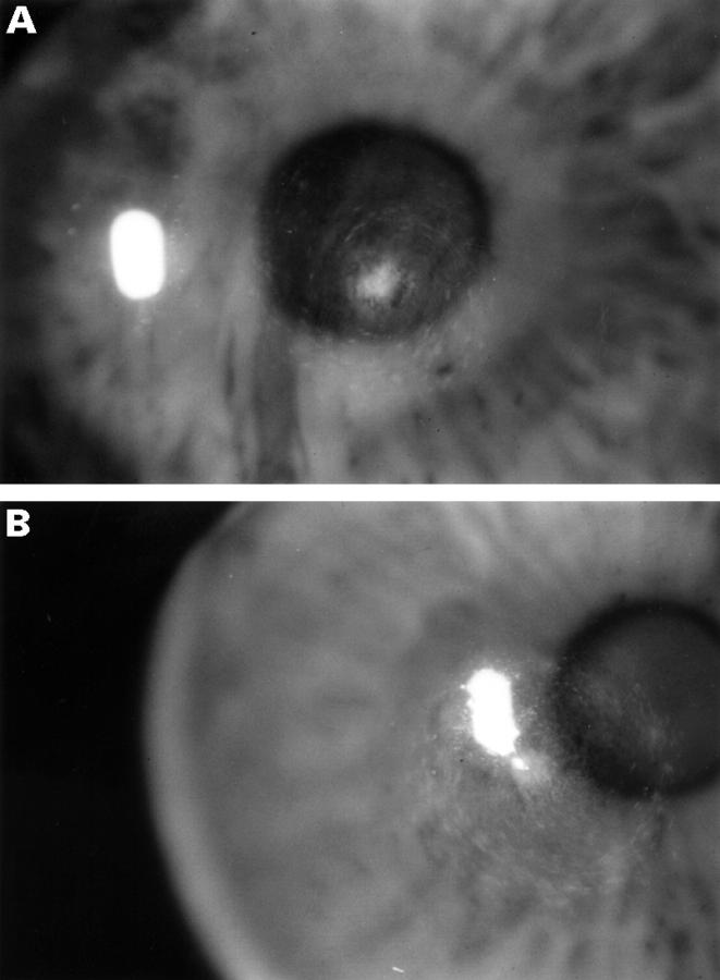
Figure 3 .
(A) A centrally located anti-clockwise whorl in the right eye of a patient with bilateral keratoconus wearing rigid gas permeable contact lenses. There is an epithelial defect at the epicentre of the whorl. (B) A centrally located clockwise whorl in the left eye of the same patient. There is a tiny epithelial defect at the epicentre of the whorl.
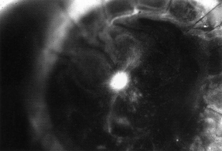
Figure 4 .
A centrally located clockwise whorl in the left eye of a patient with a corneal graft. There is an area of heaped epithelium at the centre, which is not staining with fluorescein.
Selected References
These references are in PubMed. This may not be the complete list of references from this article.
- Bron A. J. Vortex patterns of the corneal epithelium. Trans Ophthalmol Soc U K. 1973;93(0):455–472. [PubMed] [Google Scholar]
- Davanger M., Evensen A. Role of the pericorneal papillary structure in renewal of corneal epithelium. Nature. 1971 Feb 19;229(5286):560–561. doi: 10.1038/229560a0. [DOI] [PubMed] [Google Scholar]
- Dua H. S., Gomes J. A., Singh A. Corneal epithelial wound healing. Br J Ophthalmol. 1994 May;78(5):401–408. doi: 10.1136/bjo.78.5.401. [DOI] [PMC free article] [PubMed] [Google Scholar]
- Dua H. S., Singh A., Gomes J. A., Laibson P. R., Donoso L. A., Tyagi S. Vortex or whorl formation of cultured human corneal epithelial cells induced by magnetic fields. Eye (Lond) 1996;10(Pt 4):447–450. doi: 10.1038/eye.1996.98. [DOI] [PubMed] [Google Scholar]
- Dua H. S., Watson N. J., Mathur R. M., Forrester J. V. Corneal epithelial cell migration in humans: 'hurricane and blizzard keratopathy'. Eye (Lond) 1993;7(Pt 1):53–58. doi: 10.1038/eye.1993.12. [DOI] [PubMed] [Google Scholar]
- Goldberg M. F., Bron A. J. Limbal palisades of Vogt. Trans Am Ophthalmol Soc. 1982;80:155–171. [PMC free article] [PubMed] [Google Scholar]
- Lemp M. A., Mathers W. D. Corneal epithelial cell movement in humans. Eye (Lond) 1989;3(Pt 4):438–445. doi: 10.1038/eye.1989.65. [DOI] [PubMed] [Google Scholar]
- Lopatynsky M., Cohen E. J., Leavitt K. G., Laibson P. R. Corneal topography for rigid gas permeable lens fitting after penetrating keratoplasty. CLAO J. 1993 Jan;19(1):41–44. doi: 10.1097/00140068-199301000-00007. [DOI] [PubMed] [Google Scholar]
- Mathers W. D., Lemp M. A. Vortex keratopathy of the corneal graft. Cornea. 1991 Mar;10(2):93–99. doi: 10.1097/00003226-199103000-00001. [DOI] [PubMed] [Google Scholar]
- Townsend W. M. The limbal palisades of Vogt. Trans Am Ophthalmol Soc. 1991;89:721–756. [PMC free article] [PubMed] [Google Scholar]
- Tseng S. C. Concept and application of limbal stem cells. Eye (Lond) 1989;3(Pt 2):141–157. doi: 10.1038/eye.1989.22. [DOI] [PubMed] [Google Scholar]


