Abstract
AIMS—To examine the changes in the retinal nerve fibre layer (NFL) thickness with age and myopia in normal population. METHODS—Retinal nerve fibre layer thickness was measured with a scanning laser polarimeter (NFA-I) in 180 normal subjects of varying age (range 7-83 years) and in 110 eyes of 85 patients with myopia of varying degrees (range −1.00 to −15.00D). They were all voluntary Anatolian people. Superior to nasal (S/N), inferior to nasal (I/N), and the superior to inferior (S/I) ratios were used for the assessment of retinal NFL thickness. RESULTS—The mean superior NFL ratio was 2.96 and the mean inferior NFL ratio was 2.93 in normal subjects. There was a gradual decrease in NFL ratio with increasing age (simple regression analysis, p<0.05). The mean S/I ratio was 1.01 with a large variation. In patients with myopia, the mean superior NFL ratio was 2.60 and the mean inferior NFL ratio was 2.72. Superior and inferior NFL retardations, and S/I ratio in myopic patients were significantly (15.5%, 10.8%, and 4.9% respectively) lower than that of age matched normals (t test, p<0.05). There was also a gradual decrease in NFL thickness with increasing degree of myopia (simple regression analysis, p<0.05). CONCLUSIONS—Nomograms we obtained for retinal NFL thickness may serve as reference points for the assessment of normal Anatolian people and myopic patients in future studies. NFL thicknesses gradually decreased with increasing age. Patients with myopia had significantly lower NFL thicknesses than normal subjects and, although weakened by wide age range of myopic group, there is a linear relation between severity of myopia and NFL thickness in myopic patients.
Full Text
The Full Text of this article is available as a PDF (150.5 KB).
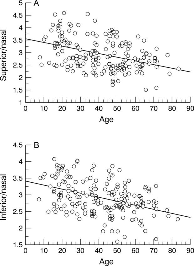
Figure 1 .

(A) Nomogram for the ratio of superior to nasal nerve fibre layer (NFL) thickness plotted against the age of the subject. Equation for the regression line: superior NFL= 3.531 − 0.014 × age R2= 0.154 (p<0.0001). (B) Nomogram for the ratio of inferior to nasal NFL thickness. Equation for the regression line: inferior NFL= 3.407 − 0.012 × age R2= 0.159 (p<0.0001).
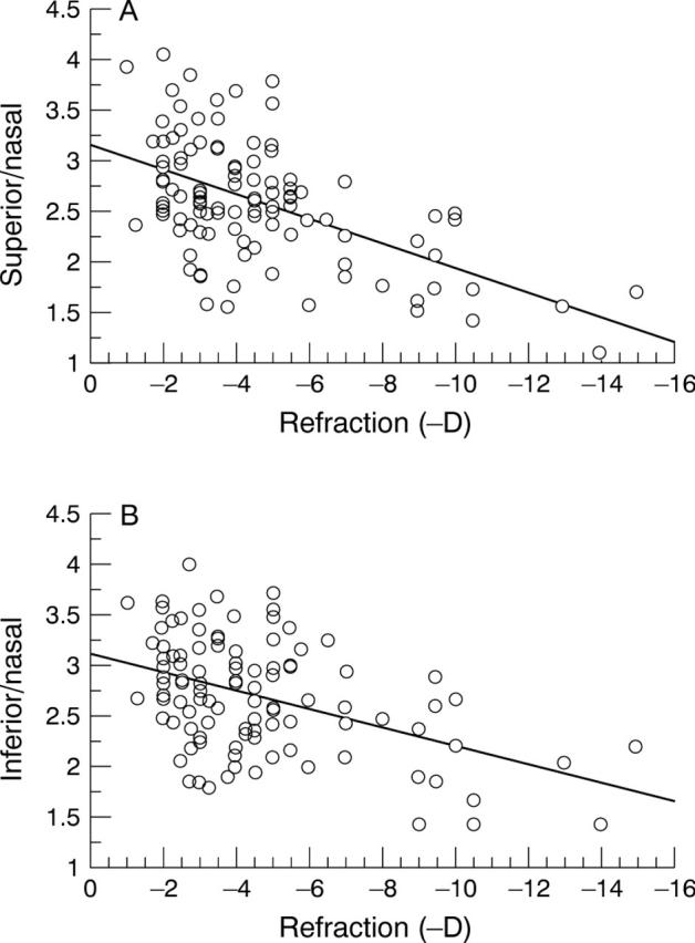
Figure 2 .

(A) Nomogram for the ratio of the superior to nasal NFL thickness plotted against the refraction (minus dioptres) of the myopic subject. Equation for the regression line: superior NFL= 3.158 − 0.122 × refraction (D) R2= 0.306 (p<0.0001). (B) Nomogram for the inferior to nasal NFL thickness plotted against the refraction of the myopic subject. Equation for the regression line: inferior NFL= 3.142 − 0.092 × refraction R2= 0.204 (p<0.0001).
Selected References
These references are in PubMed. This may not be the complete list of references from this article.
- Chen Y. F., Wang T. H., Lin L. L., Hung P. T. Influence of axial length on visual field defects in primary open-angle glaucoma. J Formos Med Assoc. 1997 Dec;96(12):968–971. [PubMed] [Google Scholar]
- Chihara E., Chihara K. Apparent cleavage of the retinal nerve fiber layer in asymptomatic eyes with high myopia. Graefes Arch Clin Exp Ophthalmol. 1992;230(5):416–420. doi: 10.1007/BF00175925. [DOI] [PubMed] [Google Scholar]
- Chihara E., Liu X., Dong J., Takashima Y., Akimoto M., Hangai M., Kuriyama S., Tanihara H., Hosoda M., Tsukahara S. Severe myopia as a risk factor for progressive visual field loss in primary open-angle glaucoma. Ophthalmologica. 1997;211(2):66–71. doi: 10.1159/000310760. [DOI] [PubMed] [Google Scholar]
- Chihara E., Sawada A. Atypical nerve fiber layer defects in high myopes with high-tension glaucoma. Arch Ophthalmol. 1990 Feb;108(2):228–232. doi: 10.1001/archopht.1990.01070040080035. [DOI] [PubMed] [Google Scholar]
- Choplin N. T., Lundy D. C., Dreher A. W. Differentiating patients with glaucoma from glaucoma suspects and normal subjects by nerve fiber layer assessment with scanning laser polarimetry. Ophthalmology. 1998 Nov;105(11):2068–2076. doi: 10.1016/S0161-6420(98)91127-7. [DOI] [PubMed] [Google Scholar]
- David R., Zangwill L. M., Tessler Z., Yassur Y. The correlation between intraocular pressure and refractive status. Arch Ophthalmol. 1985 Dec;103(12):1812–1815. doi: 10.1001/archopht.1985.01050120046017. [DOI] [PubMed] [Google Scholar]
- Ganley J. P. Epidemiological aspects of ocular hypertension. Surv Ophthalmol. 1980 Nov-Dec;25(3):130–135. doi: 10.1016/0039-6257(80)90087-9. [DOI] [PubMed] [Google Scholar]
- Hudson C. Nerve fibre layer thickness measurements derived by scanning laser polarimetry: the jury is out. Br J Ophthalmol. 1997 May;81(5):338–339. doi: 10.1136/bjo.81.5.338. [DOI] [PMC free article] [PubMed] [Google Scholar]
- Jonas J. B., Fernández M. C., Stürmer J. Pattern of glaucomatous neuroretinal rim loss. Ophthalmology. 1993 Jan;100(1):63–68. doi: 10.1016/s0161-6420(13)31694-7. [DOI] [PubMed] [Google Scholar]
- Jonas J. B., Gusek G. C., Naumann G. O. Optic disc morphometry in chronic primary open-angle glaucoma. I. Morphometric intrapapillary characteristics. Graefes Arch Clin Exp Ophthalmol. 1988;226(6):522–530. doi: 10.1007/BF02169199. [DOI] [PubMed] [Google Scholar]
- Jonas J. B., Gusek G. C., Naumann G. O. Optic disk morphometry in high myopia. Graefes Arch Clin Exp Ophthalmol. 1988;226(6):587–590. doi: 10.1007/BF02169209. [DOI] [PubMed] [Google Scholar]
- Junghardt A., Schmid M. K., Schipper I., Wildberger H., Seifert B. Reproducibility of the data determined by scanning laser polarimetry. Graefes Arch Clin Exp Ophthalmol. 1996 Oct;234(10):628–632. doi: 10.1007/BF00185296. [DOI] [PubMed] [Google Scholar]
- Pruett R. C. Progressive myopia and intraocular pressure: what is the linkage? A literature review. Acta Ophthalmol Suppl. 1988;185:117–127. doi: 10.1111/j.1755-3768.1988.tb02685.x. [DOI] [PubMed] [Google Scholar]
- Quigley H. A., Brown A. E., Morrison J. D., Drance S. M. The size and shape of the optic disc in normal human eyes. Arch Ophthalmol. 1990 Jan;108(1):51–57. doi: 10.1001/archopht.1990.01070030057028. [DOI] [PubMed] [Google Scholar]
- Tjon-Fo-Sang M. J., Lemij H. G. Retinal nerve fiber layer measurements in normal black subjects as determined with scanning laser polarimetry. Ophthalmology. 1998 Jan;105(1):78–81. doi: 10.1016/s0161-6420(98)91323-9. [DOI] [PubMed] [Google Scholar]
- Tjon-Fo-Sang M. J., de Vries J., Lemij H. G. Measurement by nerve fiber analyzer of retinal nerve fiber layer thickness in normal subjects and patients with ocular hypertension. Am J Ophthalmol. 1996 Aug;122(2):220–227. doi: 10.1016/s0002-9394(14)72013-6. [DOI] [PubMed] [Google Scholar]
- Tjon-Fo-Sang M. J., van Strik R., de Vries J., Lemij H. G. Improved reproducibility of measurements with the nerve fiber analyzer. J Glaucoma. 1997 Aug;6(4):203–211. [PubMed] [Google Scholar]
- Weinreb R. N., Dreher A. W., Coleman A., Quigley H., Shaw B., Reiter K. Histopathologic validation of Fourier-ellipsometry measurements of retinal nerve fiber layer thickness. Arch Ophthalmol. 1990 Apr;108(4):557–560. doi: 10.1001/archopht.1990.01070060105058. [DOI] [PubMed] [Google Scholar]
- Weinreb R. N., Shakiba S., Zangwill L. Scanning laser polarimetry to measure the nerve fiber layer of normal and glaucomatous eyes. Am J Ophthalmol. 1995 May;119(5):627–636. doi: 10.1016/s0002-9394(14)70221-1. [DOI] [PubMed] [Google Scholar]
- Zangwill L., Berry C. A., Garden V. S., Weinreb R. N. Reproducibility of retardation measurements with the nerve fiber analyzer II. J Glaucoma. 1997 Dec;6(6):384–389. [PubMed] [Google Scholar]


