Abstract
AIMS—To determine the prevalence of polypoidal choroidopathy in consecutive patients presenting with large haemorrhagic and exudative neurosensory retinal and retinal pigment epithelial detachments (PEDs) of over 2 mm in diameter in the absence of drusen. METHODS—40 patients were identified over a 5 month period of which 29 had haemorrhagic detachments, and 11 had purely exudative detachments. All had indocyanine green (ICG) angiography, and the presence was sought of large blood vessels in the choroid associated with localised dilated terminals that filled slowly and leaked ICG. RESULTS—In 34 cases (85%) there was an appearance consistent with previous descriptions of idiopathic polypoidal choroidal vasculopathy. Of the six without polypoidal lesions the disorder was attributed to choroidal neovascularisation in four, chorioretinitis in one, and a fibrovascular PED in one. Of those with polypoidal lesions 20 (65%) were female, the mean age was 65.4 years (range 44-88), and 25 (74%) were white, seven (20%) black, and two (6%) east Asian. Eight had a history of hypertension. Visual acuity varied from 6/6 to counting fingers in the involved eye (mean 6/24). Bilateral polypoidal choroidal lesions were demonstrated in 16 patients (47%). The predominant location for these lesions was the macular region in 23 patients (68%). Polypoidal vasculopathy was found in 16 patients (47%) who had a previous diagnosis of age related macular disease (AMD). No patients had evidence of intraocular inflammation. CONCLUSIONS—In a largely white patient population a high proportion of patients with haemorrhagic and exudative PEDs has evidence of polypoidal lesions on ICG angiography.
Full Text
The Full Text of this article is available as a PDF (219.7 KB).
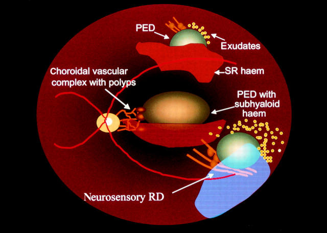
Figure 1 .
Illustration of three common presentations of idiopathic polypoidal choroidal vasculopathy. Along the superotemporal arcade, there are characteristic dilated choroidal vessels with an overlying pigment epithelial detachment (PED) and subretinal haemorrhage. Near the optic nerve there is a network of dilated choroidal vessels with polypoidal terminations with an overlying PED and subhyaloid haemorrhage. Along the inferotemporal arcade there is a ring of exudates surrounding a serous PED and neurosensory retinal detachment (RD) which overlies the polypoidal elements.
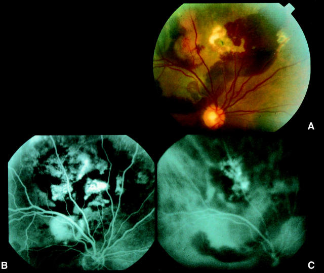
Figure 2 .
Right eye of a 68 year old black male with 6/36 vision. (A) Photograph showing a large area of subretinal haemorrhage with exudate and hyperpigmentary changes located superior to the superotemporal arcades. Along the superotemporal vessel there are two elevated subretinal lesions. (B) Fluorescein angiogram showing blockage of fluorescence by the subretinal blood and multiple areas of hyperfluorescence. (C) ICG angiogram showing a choroidal vascular network terminating in multiple polypoidal-like elements along the superior vessel which correspond to the subretinal lesions seen on the photograph.
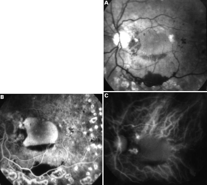
Figure 3 .
(A) Photograph of the left eye of a 75 year old black man with 6/18 vision showing a haemorrhagic PED with a nasal notch, scattered intraretinal haemorrhages, and old peripheral panretinal laser photocoagulation scars inferiorly. (B) Mid-phase fluorescein angiogram showing staining of the PED, hyperfluorescence in the area of the notch, blockage of fluorescence by the haemorrhage, and hyperfluorescent staining of the peripheral laser photocoagulation scars. (C) Early ICG angiogram showing two areas of a grape-like cluster of polyps in the peripapillary region in the area corresponding to the notch seen on fluorescein angiography.
Figure 4 .
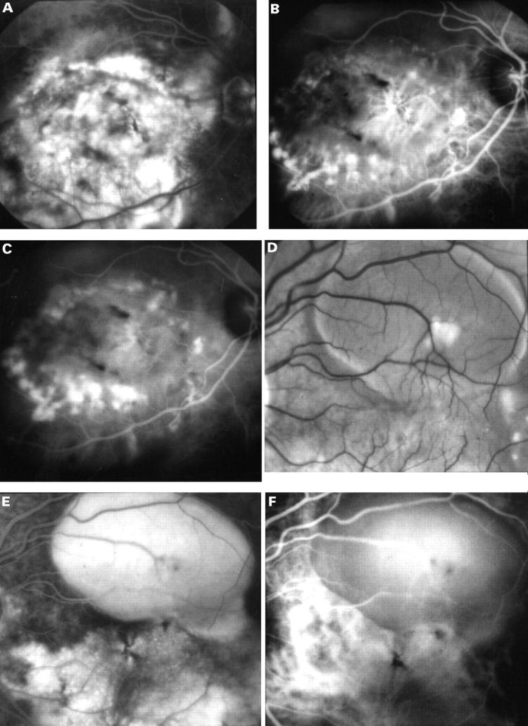
(A) A late fluorescein angiogram of the right eye of a 54 year old white man with counting fingers vision showing diffuse areas of hyperfluorescence between the temporal vascular arcades corresponding to a large serous elevation of the retina with subretinal fibrosis, hyperpigmentary changes, and subretinal and intraretinal haemorrhages. (B) Early ICG angiogram showing a ring of polyp-like aneurysmal dilatations of the choroidal vasculature in the macular region. In addition, there appears to be a choroidal neovascular complex centrally. (C) The mid-phase ICG showing staining of these polypoidal lesions. (D) In his left eye there was a pigment epithelial detachment. (E) Fluorescein angiography showed filling of the detachment and diffuse hyperfluorescence below and nasal to the lesion. (F) ICG angiography revealed a large diameter vessel complex at the same site with terminal dilatation of the vessels.
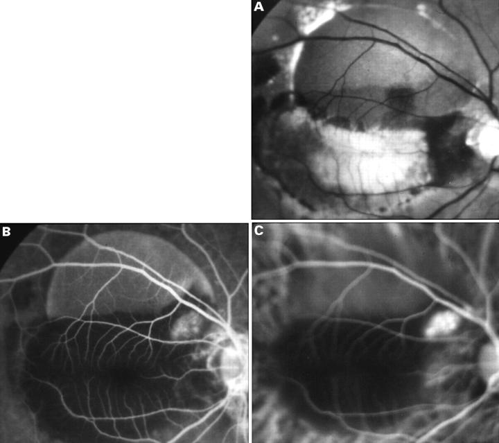
Figure 5 .
(A) The right eye of a 58 year old black male with 3/60 vision showing a large serous PED along the superotemporal arcade with surrounding subretinal haemorrhage and intraretinal exudate. (B) An early phase fluorescein angiogram showing filling of the PED, blockage of fluorescence by the subretinal exudate and haemorrhage, and a distinct hyperfluorescent lesion adjacent to the PED near the optic nerve head. (C) An early phase ICG angiogram showing a complex of polypoidal lesions in a "grape-like cluster" adjacent to the PED corresponding to the area of hyperfluorescence seen on the fluorescein angiogram.
Selected References
These references are in PubMed. This may not be the complete list of references from this article.
- Bird A. C. Doyne Lecture. Pathogenesis of retinal pigment epithelial detachment in the elderly; the relevance of Bruch's membrane change. Eye (Lond) 1991;5(Pt 1):1–12. doi: 10.1038/eye.1991.2. [DOI] [PubMed] [Google Scholar]
- Destro M., Puliafito C. A. Indocyanine green videoangiography of choroidal neovascularization. Ophthalmology. 1989 Jun;96(6):846–853. doi: 10.1016/s0161-6420(89)32826-0. [DOI] [PubMed] [Google Scholar]
- Gomez-Ulla F., Gonzalez F., Torreiro M. G. Diode laser photocoagulation in idiopathic polypoidal choroidal vasculopathy. Retina. 1998;18(5):481–483. doi: 10.1097/00006982-199805000-00021. [DOI] [PubMed] [Google Scholar]
- Green W. R., McDonnell P. J., Yeo J. H. Pathologic features of senile macular degeneration. Ophthalmology. 1985 May;92(5):615–627. [PubMed] [Google Scholar]
- Kleiner R. C., Brucker A. J., Johnston R. L. The posterior uveal bleeding syndrome. Retina. 1990;10(1):9–17. [PubMed] [Google Scholar]
- Lim J. I., Aaberg T. M., Capone A., Jr, Sternberg P., Jr Indocyanine green angiography-guided photocoagulation of choroidal neovascularization associated with retinal pigment epithelial detachment. Am J Ophthalmol. 1997 Apr;123(4):524–532. doi: 10.1016/s0002-9394(14)70178-3. [DOI] [PubMed] [Google Scholar]
- MacCumber M. W., Dastgheib K., Bressler N. M., Chan C. C., Harris M., Fine S., Green W. R. Clinicopathologic correlation of the multiple recurrent serosanguineous retinal pigment epithelial detachments syndrome. Retina. 1994;14(2):143–152. doi: 10.1097/00006982-199414020-00007. [DOI] [PubMed] [Google Scholar]
- Meredith T. A., Braley R. E., Aaberg T. M. Natural history of serous detachments of the retinal pigment epithelium. Am J Ophthalmol. 1979 Oct;88(4):643–651. doi: 10.1016/0002-9394(79)90659-7. [DOI] [PubMed] [Google Scholar]
- Moore D. J., Hussain A. A., Marshall J. Age-related variation in the hydraulic conductivity of Bruch's membrane. Invest Ophthalmol Vis Sci. 1995 Jun;36(7):1290–1297. [PubMed] [Google Scholar]
- Moorthy R. S., Lyon A. T., Rabb M. F., Spaide R. F., Yannuzzi L. A., Jampol L. M. Idiopathic polypoidal choroidal vasculopathy of the macula. Ophthalmology. 1998 Aug;105(8):1380–1385. doi: 10.1016/S0161-6420(98)98016-2. [DOI] [PubMed] [Google Scholar]
- Perkovich B. T., Zakov Z. N., Berlin L. A., Weidenthal D., Avins L. R. An update on multiple recurrent serosanguineous retinal pigment epithelial detachments in black women. Retina. 1990;10(1):18–26. [PubMed] [Google Scholar]
- Phillips W. B., 2nd, Regillo C. D., Maguire J. I. Indocyanine green angiography of idiopathic polypoidal choroidal vasculopathy. Ophthalmic Surg Lasers. 1996 Jun;27(6):467–470. [PubMed] [Google Scholar]
- Piccolino F. C. Central serous chorioretinopathy: some considerations on the pathogenesis. Ophthalmologica. 1981;182(4):204–210. doi: 10.1159/000309115. [DOI] [PubMed] [Google Scholar]
- Ross R. D., Gitter K. A., Cohen G., Schomaker K. S. Idiopathic polypoidal choroidal vasculopathy associated with retinal arterial macroaneurysm and hypertensive retinopathy. Retina. 1996;16(2):105–111. doi: 10.1097/00006982-199616020-00003. [DOI] [PubMed] [Google Scholar]
- Schneider U., Gelisken F., Kreissig I. Indocyanine green angiography and idiopathic polypoidal choroidal vasculopathy. Br J Ophthalmol. 1998 Jan;82(1):98–99. doi: 10.1136/bjo.82.1.98. [DOI] [PMC free article] [PubMed] [Google Scholar]
- Spaide R. F., Yannuzzi L. A., Slakter J. S., Sorenson J., Orlach D. A. Indocyanine green videoangiography of idiopathic polypoidal choroidal vasculopathy. Retina. 1995;15(2):100–110. doi: 10.1097/00006982-199515020-00003. [DOI] [PubMed] [Google Scholar]
- Stern R. M., Zakov Z. N., Zegarra H., Gutman F. A. Multiple recurrent serosanguineous retinal pigment epithelial detachments in black women. Am J Ophthalmol. 1985 Oct 15;100(4):560–569. doi: 10.1016/0002-9394(85)90682-8. [DOI] [PubMed] [Google Scholar]
- Yannuzzi L. A., Ciardella A., Spaide R. F., Rabb M., Freund K. B., Orlock D. A. The expanding clinical spectrum of idiopathic polypoidal choroidal vasculopathy. Arch Ophthalmol. 1997 Apr;115(4):478–485. doi: 10.1001/archopht.1997.01100150480005. [DOI] [PubMed] [Google Scholar]
- Yannuzzi L. A., Hope-Ross M., Slakter J. S., Guyer D. R., Sorenson J. A., Ho A. C., Sperber D. E., Freund K. B., Orlock D. A. Analysis of vascularized pigment epithelial detachments using indocyanine green videoangiography. Retina. 1994;14(2):99–113. doi: 10.1097/00006982-199414020-00003. [DOI] [PubMed] [Google Scholar]
- Yannuzzi L. A., Nogueira F. B., Spaide R. F., Guyer D. R., Orlock D. A., Colombero D., Freund K. B. Idiopathic polypoidal choroidal vasculopathy: a peripheral lesion. Arch Ophthalmol. 1998 Mar;116(3):382–383. [PubMed] [Google Scholar]
- Yannuzzi L. A., Sorenson J., Spaide R. F., Lipson B. Idiopathic polypoidal choroidal vasculopathy (IPCV). Retina. 1990;10(1):1–8. [PubMed] [Google Scholar]


