Full Text
The Full Text of this article is available as a PDF (324.8 KB).
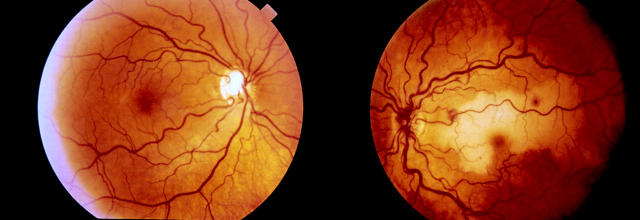
Figure 1 .
(Left) The right fundus at presentation showing dilated retinal veins and a pathologically cupped optic disc. (Right) The left fundus at presentation showing a swollen, haemorrhagic optic disc, dilated veins in all four quadrants and scattered perivenous retinal haemorrhages. There is retinal whitening corresponding with the distribution of a cilioretinal artery.
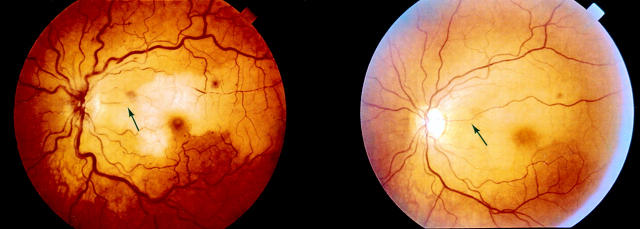
Figure 2 .
(Left) The left fundus 1 week after presentation. The cilioretinal artery supplying the superior macula is narrow and irregular in calibre with fractionation of the blood column (arrow). (Right) The left fundus 2 months after presentation. The calibre of the cilioretinal artery has returned to normal (arrow) and most retinal haemorrhages have resolved.


