Abstract
BACKGROUND—Spontaneous bullous serous retinal detachment (RD) with subretinal exudation complicating idiopathic central serous chorioretinopathy (ICSC) is a rare and infrequently described clinical entity. Clinical observations are described on this variant form in 11 patients, the largest series reported to date. METHODS—13 eyes of 11 Indian patients having this entity were followed up clinically and angiographically for 12-24 months (retrospective, longitudinal). None of the patients had any previous history of other diseases nor were they on any medications. Four eyes received laser treatment (group A); nine eyes were not treated (group B). RESULTS—All 11 patients were male, aged 23-49 years (median 37 years). The clinical and photographic records revealed subretinal exudation and inferior bullous serous RD complicating ICSC with evidence of large, single or multiple, leaking retinal pigment epithelial detachments (PEDs) in all the cases. In group A, resolution of serous RD occurred in 12 weeks (median) with a visual recovery of ⩾20/30 in three out of four eyes while in group B resolution of serous retinal detachment was observed in 14 weeks (median) with eight out of nine eyes achieving a visual acuity of ⩾20/30. Subretinal fibrosis developed in two eyes in group A and none of the eyes in group B. CONCLUSION—The disease is an exaggerated form of ICSC and can occur spontaneously without any history of corticosteroid therapy. Recognition of this atypical presentation is important to avoid inappropriate treatment. These observations suggest that with respect to the duration of the disease and the final visual outcome laser therapy offers no additional benefit over the natural course of this variant form of ICSC.
Full Text
The Full Text of this article is available as a PDF (197.1 KB).
Figure 1 .
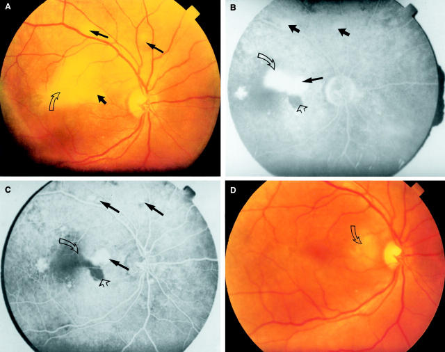
Case 4 right eye. (A) Fundus appearance at presentation showing two pigment epithelium detachments (PEDs) outside the superior temporal arcade (thin solid arrows). Notice a subtle PED nasal to the fovea (thick solid arrow) in the vicinity of subretinal exudation (open arrow). (B) Midphase fluorescein angiogram demonstrating multiple PEDs (solid arrows). Hypofluorescent areas are due to subretinal exudation (open arrow) and a hidden PED underneath the subretinal exudation (open arrowhead). (C) Late phase fluorescein angiogram showing multiple PEDs (thick solid arrows), a leaking PED (thin solid arrow), staining of subretinal exudation (open arrow), and persistent hypofluorescence due to a PED (open arrowhead) hidden under subretinal exudation. (D) Fundus appearance 14 weeks after onset (non-laser treated) showing near total resolution of subretinal exudation temporal to the disc (open arrow).
Figure 2 .

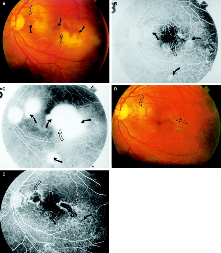
Case 4 left eye. (A) Fundus appearance at presentation demonstrating massive subretinal exudation and serous retinal detachment in the posterior pole. Notice hidden PEDs (solid arrows) in the vicinity of subretinal exudation (open arrows). (B) Midphase fluorescein angiogram demonstrating multiple PEDs (solid arrows), and hypofluorescent areas are due to a hidden PED (open arrowhead) and subretinal exudation (open arrow). (C) Late phase fluorescein angiogram showing leakage from the edges of PEDs (solid arrows), and staining of the subretinal exudation (open arrow). (D) Fundus appearance 12 weeks after laser photocoagulation showing subretinal fibrotic membrane around the fovea (open arrows). (E) Late phase angiogram 16 weeks after laser photocoagulation showing substantial amount of RPE degeneration (solid arrows) and subretinal fibrotic membranes (open arrows).
Selected References
These references are in PubMed. This may not be the complete list of references from this article.
- Akiyama K., Kawamura M., Ogata T., Tanaka E. Retinal vascular loss in idiopathic central serous chorioretinopathy with bullous retinal detachment. Ophthalmology. 1987 Dec;94(12):1605–1609. doi: 10.1016/s0161-6420(87)33243-9. [DOI] [PubMed] [Google Scholar]
- Dellaporta A. Central serous retinopathy. Trans Am Ophthalmol Soc. 1976;74:144–153. [PMC free article] [PubMed] [Google Scholar]
- Desatnik H. R., Gutman F. A. Bilateral exudative retinal detachment complicating systemic corticosteroid therapy in the presence of renal failure. Am J Ophthalmol. 1996 Sep;122(3):432–434. doi: 10.1016/s0002-9394(14)72075-6. [DOI] [PubMed] [Google Scholar]
- Ficker L., Vafidis G., While A., Leaver P. Long-term follow-up of a prospective trial of argon laser photocoagulation in the treatment of central serous retinopathy. Br J Ophthalmol. 1988 Nov;72(11):829–834. doi: 10.1136/bjo.72.11.829. [DOI] [PMC free article] [PubMed] [Google Scholar]
- Friberg T. R., Eller A. W. Serous retinal detachment resembling central serous chorioretinopathy following organ transplantation. Graefes Arch Clin Exp Ophthalmol. 1990;228(4):305–309. doi: 10.1007/BF00920052. [DOI] [PubMed] [Google Scholar]
- Gass J. D. Bullous retinal detachment and multiple retinal pigment epithelial detachments in patients receiving hemodialysis. Graefes Arch Clin Exp Ophthalmol. 1992;230(5):454–458. doi: 10.1007/BF00175933. [DOI] [PubMed] [Google Scholar]
- Gass J. D. Bullous retinal detachment. An unusual manifestation of idiopathic central serous choroidopathy. Am J Ophthalmol. 1973 May;75(5):810–821. doi: 10.1016/0002-9394(73)90887-8. [DOI] [PubMed] [Google Scholar]
- Gass J. D. Central serous chorioretinopathy and white subretinal exudation during pregnancy. Arch Ophthalmol. 1991 May;109(5):677–681. doi: 10.1001/archopht.1991.01080050091036. [DOI] [PubMed] [Google Scholar]
- Gass J. D., Little H. Bilateral bullous exudative retinal detachment complicating idiopathic central serous chorioretinopathy during systemic corticosteroid therapy. Ophthalmology. 1995 May;102(5):737–747. doi: 10.1016/s0161-6420(95)30960-8. [DOI] [PubMed] [Google Scholar]
- Gass J. D., Slamovits T. L., Fuller D. G., Gieser R. G., Lean J. S. Posterior chorioretinopathy and retinal detachment after organ transplantation. Arch Ophthalmol. 1992 Dec;110(12):1717–1722. doi: 10.1001/archopht.1992.01080240057030. [DOI] [PubMed] [Google Scholar]
- Gelber G. S., Schatz H. Loss of vision due to central serous chorioretinopathy following psychological stress. Am J Psychiatry. 1987 Jan;144(1):46–50. doi: 10.1176/ajp.144.1.46. [DOI] [PubMed] [Google Scholar]
- Jabs D. A., Hanneken A. M., Schachat A. P., Fine S. L. Choroidopathy in systemic lupus erythematosus. Arch Ophthalmol. 1988 Feb;106(2):230–234. doi: 10.1001/archopht.1988.01060130240036. [DOI] [PubMed] [Google Scholar]
- KLIEN B. A. Macular lesions of vascular origin. II. Functional vascular conditions leading to damage of the macula lutea. Am J Ophthalmol. 1953 Jan;36(1):1–13. [PubMed] [Google Scholar]
- Klein M. L., Van Buskirk E. M., Friedman E., Gragoudas E., Chandra S. Experience with nontreatment of central serous choroidopathy. Arch Ophthalmol. 1974 Apr;91(4):247–250. doi: 10.1001/archopht.1974.03900060257001. [DOI] [PubMed] [Google Scholar]
- Laatikainen L. Diffuse chronic retinal pigment epitheliopathy and exudative retinal detachment. Acta Ophthalmol (Copenh) 1994 Oct;72(5):533–536. doi: 10.1111/j.1755-3768.1994.tb07175.x. [DOI] [PubMed] [Google Scholar]
- Leaver P., Williams C. Argon laser photocoagulation in the treatment of central serous retinopathy. Br J Ophthalmol. 1979 Oct;63(10):674–677. doi: 10.1136/bjo.63.10.674. [DOI] [PMC free article] [PubMed] [Google Scholar]
- Matsuo T., Nakayama T., Koyama T., Matsuo N. Multifocal pigment epithelial damages with serous retinal detachment in systemic lupus erythematosus. Ophthalmologica. 1987;195(2):97–102. doi: 10.1159/000309795. [DOI] [PubMed] [Google Scholar]
- Mazzuca D. E., Benson W. E. Central serous retinopathy: variants. Surv Ophthalmol. 1986 Nov-Dec;31(3):170–174. doi: 10.1016/0039-6257(86)90036-6. [DOI] [PubMed] [Google Scholar]
- Novak M. A., Singerman L. J., Rice T. A. Krypton and argon laser photocoagulation for central serous chorioretinopathy. Retina. 1987 Fall;7(3):162–169. doi: 10.1097/00006982-198700730-00005. [DOI] [PubMed] [Google Scholar]
- Quillen D. A., Gass D. M., Brod R. D., Gardner T. W., Blankenship G. W., Gottlieb J. L. Central serous chorioretinopathy in women. Ophthalmology. 1996 Jan;103(1):72–79. doi: 10.1016/s0161-6420(96)30730-6. [DOI] [PubMed] [Google Scholar]
- Robertson D. M., Ilstrup D. Direct, indirect, and sham laser photocoagulation in the management of central serous chorioretinopathy. Am J Ophthalmol. 1983 Apr;95(4):457–466. doi: 10.1016/0002-9394(83)90265-9. [DOI] [PubMed] [Google Scholar]
- Schatz H., McDonald H. R., Johnson R. N., Chan C. K., Irvine A. R., Berger A. R., Folk J. C., Robertson D. M. Subretinal fibrosis in central serous chorioretinopathy. Ophthalmology. 1995 Jul;102(7):1077–1088. doi: 10.1016/s0161-6420(95)30908-6. [DOI] [PubMed] [Google Scholar]
- Slusher M. M. Krypton red laser photocoagulation in selected cases of central serous chorioretinopathy. Retina. 1986 Spring-Summer;6(2):81–84. doi: 10.1097/00006982-198600620-00003. [DOI] [PubMed] [Google Scholar]
- Tsukahara I., Uyama M. Central serous choroidopathy with bullous retinal detachment. Albrecht Von Graefes Arch Klin Exp Ophthalmol. 1978 May 16;206(3):169–178. doi: 10.1007/BF00414743. [DOI] [PubMed] [Google Scholar]
- Wakakura M., Ishikawa S. Central serous chorioretinopathy complicating systemic corticosteroid treatment. Br J Ophthalmol. 1984 May;68(5):329–331. doi: 10.1136/bjo.68.5.329. [DOI] [PMC free article] [PubMed] [Google Scholar]
- Yannuzzi L. A. Type-A behavior and central serous chorioretinopathy. Retina. 1987 Summer;7(2):111–131. doi: 10.1097/00006982-198700720-00009. [DOI] [PubMed] [Google Scholar]
- Yoshioka H., Katsume Y., Akune H. Experimental central serous chorioretinopathy in monkey eyes: fluorescein angiographic findings. Ophthalmologica. 1982;185(3):168–178. doi: 10.1159/000309239. [DOI] [PubMed] [Google Scholar]


