Full Text
The Full Text of this article is available as a PDF (324.7 KB).
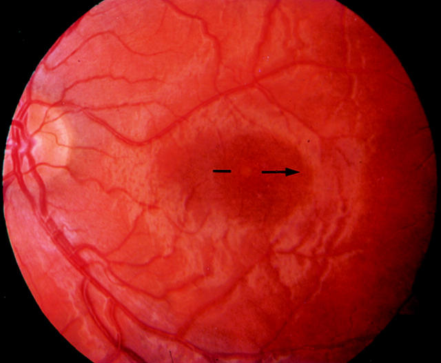
Figure 1 .
Acute stage of solar retinopathy showing a yellow lesion surrounded by a circular red area (patient 1, left eye).
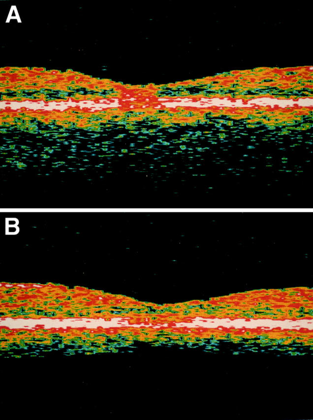
Figure 2 .

(A) OCT examination of the same eye 48 hours after exposure revealed a hyperreflective area in the fovea affecting all retinal layers. No increase in retinal thickness could be demonstrated. The location of the OCT scan is shown on the corresponding fundus photograph (Fig 1). (B) OCT examination 9 days after exposure. The hyperreflective area in the fovea was no longer visible, whereas an increasing alteration of the RPE and choriocapillary layer could be demonstrated. Visual acuity increased from 0.1 to 0.16. Still no change in retinal thickness occurred.


