Abstract
AIM—To compare the effect of altitudinal asymmetric glaucomatous damage on retinal microcirculation in patients with normal pressure glaucoma (NPG). METHODS—In a prospective cross sectional study patients with NPG (washed out for antiglaucomatous therapy) and altitudinal asymmetric perimetric findings between the superior and inferior hemisphere (Humphrey 24-2) (n=18) were included and compared with 20 NPG patients with symmetrical field defects and 18 healthy subjects. Fluorescein angiograms were performed using a scanning laser ophthalmoscope. Using digital image analysis, arteriovenous passage time (AVP) and vessel diameters were assessed for comparison of corresponding affected and less affected temporal arcades. RESULTS—Both affected and less affected hemispheres showed significantly prolonged AVP times (p<0.001) when compared with healthy subject data. In hemispheres with more severe glaucomatous field loss the AVP times were significantly (p=0.04) prolonged compared with the less affected hemisphere (AVP affected 3.1 (SD 7) seconds v AVP less affected 2.61 (1.4) seconds). There was no asymmetry effect on arterial and venous diameter measurements. CONCLUSION—Altitudinal visual field defects are linked together with circulatory deficits of the retinal tissue. The attenuated circulation seems to be a considerable factor in the natural course of glaucomatous optic neuropathy.
Full Text
The Full Text of this article is available as a PDF (188.5 KB).
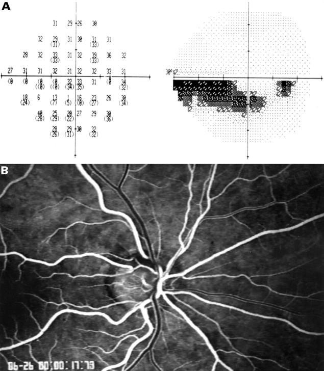
Figure 1 .
(A) Asymmetric visual field loss of the inferior visual field (Humphrey 24-2) with an arcuate scotoma in the inferior hemifield. (B) Corresponding fluorescein angiogram with focal ischaemia of the optic disc temporally superior (AVP time of the affected temporal superior vessel: 2.17 seconds ; the less affected temporal inferior vessel formation: 1.67 seconds).
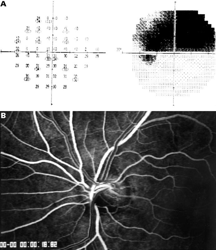
Figure 2 .
(A) Asymmetric hemifield visual field loss of the superior visual field (Humphrey 24-2). (B) Corresponding fluorescein angiogram with ischaemia of the inferior portion of the optic disc (AVP of affected inferior vessel formation: 3.6 seconds; AVP of less affected superior vessel formation: 2.23 seconds). Note the visible different fluorescein intensity of the laminar stream of the dye in the veins superiorly and inferiorly.
Selected References
These references are in PubMed. This may not be the complete list of references from this article.
- Anderson D. R. Glaucoma, capillaries and pericytes. 1. Blood flow regulation. Ophthalmologica. 1996;210(5):257–262. doi: 10.1159/000310722. [DOI] [PubMed] [Google Scholar]
- Bennett A. G., Rudnicka A. R., Edgar D. F. Improvements on Littmann's method of determining the size of retinal features by fundus photography. Graefes Arch Clin Exp Ophthalmol. 1994 Jun;232(6):361–367. doi: 10.1007/BF00175988. [DOI] [PubMed] [Google Scholar]
- Bertram B., Wolf S., Fiehöfer S., Schulte K., Arend O., Reim M. Retinal circulation times in diabetes mellitus type 1. Br J Ophthalmol. 1991 Aug;75(8):462–465. doi: 10.1136/bjo.75.8.462. [DOI] [PMC free article] [PubMed] [Google Scholar]
- Caprioli J. Neuroprotection of the optic nerve in glaucoma. Acta Ophthalmol Scand. 1997 Aug;75(4):364–367. doi: 10.1111/j.1600-0420.1997.tb00391.x. [DOI] [PubMed] [Google Scholar]
- Chylack L. T., Jr, Wolfe J. K., Singer D. M., Leske M. C., Bullimore M. A., Bailey I. L., Friend J., McCarthy D., Wu S. Y. The Lens Opacities Classification System III. The Longitudinal Study of Cataract Study Group. Arch Ophthalmol. 1993 Jun;111(6):831–836. doi: 10.1001/archopht.1993.01090060119035. [DOI] [PubMed] [Google Scholar]
- Delori F. C., Fitch K. A., Feke G. T., Deupree D. M., Weiter J. J. Evaluation of micrometric and microdensitometric methods for measuring the width of retinal vessel images on fundus photographs. Graefes Arch Clin Exp Ophthalmol. 1988;226(4):393–399. doi: 10.1007/BF02172974. [DOI] [PubMed] [Google Scholar]
- Drance S. M., Douglas G. R., Wijsman K., Schulzer M., Britton R. J. Response of blood flow to warm and cold in normal and low-tension glaucoma patients. Am J Ophthalmol. 1988 Jan 15;105(1):35–39. doi: 10.1016/0002-9394(88)90118-3. [DOI] [PubMed] [Google Scholar]
- Fontana L., Poinoosawmy D., Bunce C. V., O'Brien C., Hitchings R. A. Pulsatile ocular blood flow investigation in asymmetric normal tension glaucoma and normal subjects. Br J Ophthalmol. 1998 Jul;82(7):731–736. doi: 10.1136/bjo.82.7.731. [DOI] [PMC free article] [PubMed] [Google Scholar]
- Frisén L., Frisén M. A simple relationship between the probability distribution of visual acuity and the density of retinal output channels. Acta Ophthalmol (Copenh) 1976 Aug;54(4):437–444. doi: 10.1111/j.1755-3768.1976.tb01275.x. [DOI] [PubMed] [Google Scholar]
- Grunwald J. E., Riva C. E., Stone R. A., Keates E. U., Petrig B. L. Retinal autoregulation in open-angle glaucoma. Ophthalmology. 1984 Dec;91(12):1690–1694. doi: 10.1016/s0161-6420(84)34091-x. [DOI] [PubMed] [Google Scholar]
- Jonas J. B., Gründler A. E., Gonzales-Cortés J. Pressure-dependent neuroretinal rim loss in normal-pressure glaucoma. Am J Ophthalmol. 1998 Feb;125(2):137–144. doi: 10.1016/s0002-9394(99)80083-x. [DOI] [PubMed] [Google Scholar]
- Jonas J. B., Nguyen X. N., Naumann G. O. Parapapillary retinal vessel diameter in normal and glaucoma eyes. I. Morphometric data. Invest Ophthalmol Vis Sci. 1989 Jul;30(7):1599–1603. [PubMed] [Google Scholar]
- Littmann H. Zur Bestimmung der wahren Grösse eines Objektes auf dem Hintergrund eines lebenden Auges. Klin Monbl Augenheilkd. 1988 Jan;192(1):66–67. doi: 10.1055/s-2008-1050076. [DOI] [PubMed] [Google Scholar]
- Michelson G., Langhans M. J., Harazny J., Dichtl A. Visual field defect and perfusion of the juxtapapillary retina and the neuroretinal rim area in primary open-angle glaucoma. Graefes Arch Clin Exp Ophthalmol. 1998 Feb;236(2):80–85. doi: 10.1007/s004170050046. [DOI] [PubMed] [Google Scholar]
- Nicolela M. T., Drance S. M., Rankin S. J., Buckley A. R., Walman B. E. Color Doppler imaging in patients with asymmetric glaucoma and unilateral visual field loss. Am J Ophthalmol. 1996 May;121(5):502–510. doi: 10.1016/s0002-9394(14)75424-8. [DOI] [PubMed] [Google Scholar]
- Phelps C. D., Corbett J. J. Migraine and low-tension glaucoma. A case-control study. Invest Ophthalmol Vis Sci. 1985 Aug;26(8):1105–1108. [PubMed] [Google Scholar]
- Poinoosawmy D., Fontana L., Wu J. X., Bunce C. V., Hitchings R. A. Frequency of asymmetric visual field defects in normal-tension and high-tension glaucoma. Ophthalmology. 1998 Jun;105(6):988–991. doi: 10.1016/S0161-6420(98)96049-3. [DOI] [PubMed] [Google Scholar]
- Quigley H. A., Addicks E. M. Regional differences in the structure of the lamina cribrosa and their relation to glaucomatous optic nerve damage. Arch Ophthalmol. 1981 Jan;99(1):137–143. doi: 10.1001/archopht.1981.03930010139020. [DOI] [PubMed] [Google Scholar]
- Rankin S. J., Walman B. E., Buckley A. R., Drance S. M. Color Doppler imaging and spectral analysis of the optic nerve vasculature in glaucoma. Am J Ophthalmol. 1995 Jun;119(6):685–693. doi: 10.1016/s0002-9394(14)72771-0. [DOI] [PubMed] [Google Scholar]
- Remky A., Arend O., Beausencourt E., Elsner A. E., Bertram B. Retinale Gefässe vor und nach Photokoagulation bei diabetischer Retinopathie. Durchmesserbestimmungen mittels digitalisierter Farbdiapositive. Klin Monbl Augenheilkd. 1996 Aug-Sep;209(2-3):79–83. doi: 10.1055/s-2008-1035282. [DOI] [PubMed] [Google Scholar]
- Richard G., Hackelbusch R., Schmidt K. U., Schäfer M. Untersuchung zur Haemodynamik des Auges bei Glaucoma chronicum simplex und low tension Glaucom--eine videoangiographische Studie. Fortschr Ophthalmol. 1988;85(4):369–372. [PubMed] [Google Scholar]
- Rojanapongpun P., Drance S. M., Morrison B. J. Ophthalmic artery flow velocity in glaucomatous and normal subjects. Br J Ophthalmol. 1993 Jan;77(1):25–29. doi: 10.1136/bjo.77.1.25. [DOI] [PMC free article] [PubMed] [Google Scholar]
- Siegner S. W., Netland P. A. Optic disc hemorrhages and progression of glaucoma. Ophthalmology. 1996 Jul;103(7):1014–1024. doi: 10.1016/s0161-6420(96)30572-1. [DOI] [PubMed] [Google Scholar]
- Spaeth G. L. Fluorescein angiography: its contributions towards understanding the mechanisms of visual loss in glaucoma. Trans Am Ophthalmol Soc. 1975;73:491–553. [PMC free article] [PubMed] [Google Scholar]
- Sponsel W. E., DePaul K. L., Kaufman P. L. Correlation of visual function and retinal leukocyte velocity in glaucoma. Am J Ophthalmol. 1990 Jan 15;109(1):49–54. doi: 10.1016/s0002-9394(14)75578-3. [DOI] [PubMed] [Google Scholar]
- Suzuki R., Nakayama M., Yoshino H., Kurimoto S. Comparison of laser trabeculostimulation with laser trabeculoplasty in open-angle glaucoma. Ann Ophthalmol. 1992 Jul;24(7):245–249. [PubMed] [Google Scholar]
- Vécsei P. V., Hommer A., Reitner A., Kircher K., Egger S., Schneider B., Bettelheim H. C. Farbduplex der retrobulbären Arterien bei Normaldruck- und Offenwinkelglaukom. Klin Monbl Augenheilkd. 1998 Jun;212(6):444–448. [PubMed] [Google Scholar]
- Wolf S., Arend O., Haase A., Schulte K., Remky A., Reim M. Retinal hemodynamics in patients with chronic open-angle glaucoma. Ger J Ophthalmol. 1995 Sep;4(5):279–282. [PubMed] [Google Scholar]
- Wolf S., Arend O., Reim M. Measurement of retinal hemodynamics with scanning laser ophthalmoscopy: reference values and variation. Surv Ophthalmol. 1994 May;38 (Suppl):S95–100. doi: 10.1016/0039-6257(94)90052-3. [DOI] [PubMed] [Google Scholar]
- Wolf S., Arend O., Sponsel W. E., Schulte K., Cantor L. B., Reim M. Retinal hemodynamics using scanning laser ophthalmoscopy and hemorheology in chronic open-angle glaucoma. Ophthalmology. 1993 Oct;100(10):1561–1566. doi: 10.1016/s0161-6420(93)31444-2. [DOI] [PubMed] [Google Scholar]
- Yamazaki Y., Drance S. M. The relationship between progression of visual field defects and retrobulbar circulation in patients with glaucoma. Am J Ophthalmol. 1997 Sep;124(3):287–295. doi: 10.1016/s0002-9394(14)70820-7. [DOI] [PubMed] [Google Scholar]


