

Full Text
The Full Text of this article is available as a PDF (381.2 KB).
Figure 1 .
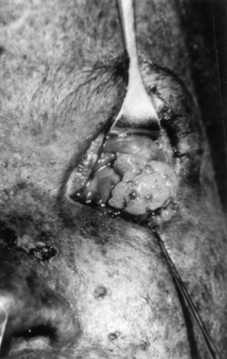
Left eye of the proband demonstrating the large pink, friable conjunctival lesion, a biopsy of which showed moderately differentiated squamous cell carcinoma. Note the scaly nature of the surrounding facial skin with actinic keratotic lesions, hypopigmented and hyperpigmented areas and crusted ulceration of the nasal bridge, all typical cutaneous lesions in xeroderma pigmentosum.
Figure 2 .
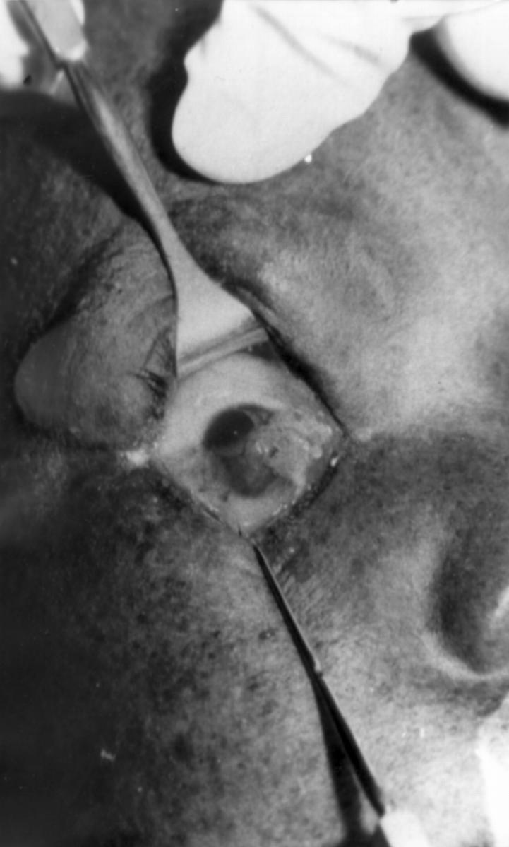
Case 3, right eye showing vascular, fleshy conjunctival tissues, a biopsy of which revealed features consistent with moderately differentiated squamous cell carcinoma. The facial skin demonstrates actinic keratotic lesions typical of xeroderma pigmentosum.