Abstract
BACKGROUND/AIMS—Deturgescence of the corneal stroma is controlled by the pumping action of the endothelial layer and can be monitored by measurement of central corneal thickness (pachymetry). Loss or damage of endothelial cells leads to an increase in corneal thickness, which may ultimately induce corneal decompensation and loss of vision. Little is known about the effect of moderate reductions in endothelial cell number on the thickness of the corneal stroma. This study aimed to investigate this matter further using patients who had incurred moderate decreases in their endothelial cell counts as a result of cataract surgery. METHODS—Central corneal thickness was measured 1 day before surgery, 1 day after surgery, and again at 3 months or 1 year. Endothelial cell counts were also performed 1 day before surgery and thereafter at 3 months or 1 year after surgery. The relationship between these two parameters was assessed statistically. Precise measurements of central corneal thickness were made by optical low coherence reflectometry. For comparative purposes, this parameter was also determined by ultrasonic pachymetry. Central corneal endothelial cell numerical density was estimated on photomicrographs taken with a specular microscope. RESULTS—All patients had significant postoperative corneal swelling on the day after surgery; preoperative values were restored by 3 and 12 months, even though significant endothelial cell losses had occurred. No correlation existed between central corneal thickness and central corneal endothelial cell numerical density. Measurements estimated by ultrasonic pachymetry were more variable and significantly higher than those determined by optical low coherence reflectometry. CONCLUSION—As long as the numerical density of the corneal endothelial cells does not fall below the physiological threshold, a moderate decrease in this parameter does not compromise the pumping activity of the layer as a whole.
Full Text
The Full Text of this article is available as a PDF (121.5 KB).
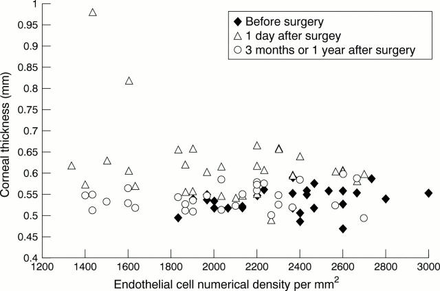
Figure 1 .
Corneal thickness and endothelial cell numerical density measured by optical low coherence reflectometry 1 day before surgery, 1 day after surgery, and 3 or 12 months after surgery.
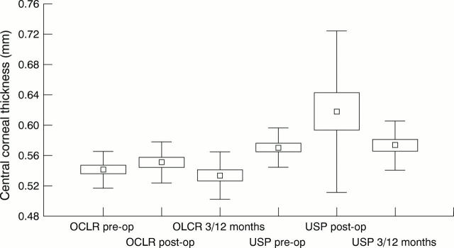
Figure 2 .
Central corneal thickness measured by optical low coherence reflectometry (OLCR) and ultrasonic pachymetry (USP) 1 day before surgery (pre-op), 1 day after surgery (post-op), and 3 or 12 months after surgery.
Selected References
These references are in PubMed. This may not be the complete list of references from this article.
- Amon M., Menapace R., Radax U., Papapanos P. Endothelial cell density and corneal pachometry after no-stitch, small-incision cataract surgery. Doc Ophthalmol. 1992;81(3):301–307. doi: 10.1007/BF00161768. [DOI] [PubMed] [Google Scholar]
- Autzen T., Bjørnstrøm L. Central corneal thickness in premature babies. Acta Ophthalmol (Copenh) 1991 Apr;69(2):251–252. doi: 10.1111/j.1755-3768.1991.tb02720.x. [DOI] [PubMed] [Google Scholar]
- Bourne W. M., Kaufman H. E. Specular microscopy of human corneal endothelium in vivo. Am J Ophthalmol. 1976 Mar;81(3):319–323. doi: 10.1016/0002-9394(76)90247-6. [DOI] [PubMed] [Google Scholar]
- Bourne W. M., O'Fallon W. M. Endothelial cell loss during penetrating keratoplasty. Am J Ophthalmol. 1978 Jun;85(6):760–766. doi: 10.1016/s0002-9394(14)78102-4. [DOI] [PubMed] [Google Scholar]
- Burns R. R., Bourne W. M., Brubaker R. F. Endothelial function in patients with cornea guttata. Invest Ophthalmol Vis Sci. 1981 Jan;20(1):77–85. [PubMed] [Google Scholar]
- Böhnke M., Chavanne P., Gianotti R., Salathé R. P. High-precision, high-speed measurement of excimer laser keratectomies with a new optical pachymeter. Ger J Ophthalmol. 1996 Nov;5(6):338–342. [PubMed] [Google Scholar]
- Capella J. A. Regeneration of endothelium in diseased and injured corneas. Am J Ophthalmol. 1972 Nov;74(5):810–817. doi: 10.1016/0002-9394(72)91200-7. [DOI] [PubMed] [Google Scholar]
- Cheng H., Bates A. K., Wood L., McPherson K. Positive correlation of corneal thickness and endothelial cell loss. Serial measurements after cataract surgery. Arch Ophthalmol. 1988 Jul;106(7):920–922. doi: 10.1001/archopht.1988.01060140066026. [DOI] [PubMed] [Google Scholar]
- Ehlers N., Sorensen T., Bramsen T., Poulsen E. H. Central corneal thickness in newborns and children. Acta Ophthalmol (Copenh) 1976 Jul;54(3):285–290. doi: 10.1111/j.1755-3768.1976.tb01257.x. [DOI] [PubMed] [Google Scholar]
- Fischbarg J., Lim J. J. Role of cations, anions and carbonic anhydrase in fluid transport across rabbit corneal endothelium. J Physiol. 1974 Sep;241(3):647–675. doi: 10.1113/jphysiol.1974.sp010676. [DOI] [PMC free article] [PubMed] [Google Scholar]
- Geroski D. H., Matsuda M., Yee R. W., Edelhauser H. F. Pump function of the human corneal endothelium. Effects of age and cornea guttata. Ophthalmology. 1985 Jun;92(6):759–763. doi: 10.1016/s0161-6420(85)33973-8. [DOI] [PubMed] [Google Scholar]
- Hara T., Hara T. Postoperative change in the corneal thickness of the pseudophakic eye: amplified diurnal variation and consensual increase. J Cataract Refract Surg. 1987 May;13(3):325–329. doi: 10.1016/s0886-3350(87)80082-2. [DOI] [PubMed] [Google Scholar]
- Harper C. L., Boulton M. E., Bennett D., Marcyniuk B., Jarvis-Evans J. H., Tullo A. B., Ridgway A. E. Diurnal variations in human corneal thickness. Br J Ophthalmol. 1996 Dec;80(12):1068–1072. doi: 10.1136/bjo.80.12.1068. [DOI] [PMC free article] [PubMed] [Google Scholar]
- Herse P., Yao W. Variation of corneal thickness with age in young New Zealanders. Acta Ophthalmol (Copenh) 1993 Jun;71(3):360–364. doi: 10.1111/j.1755-3768.1993.tb07148.x. [DOI] [PubMed] [Google Scholar]
- Hodson S. Evidence for a bicarbonate-dependent sodium pump in corneal endothelium. Exp Eye Res. 1971 Jan;11(1):20–29. doi: 10.1016/s0014-4835(71)80060-x. [DOI] [PubMed] [Google Scholar]
- Kaufman H. E., Capella J. A., Robbins J. E. The human corneal endothelium. Am J Ophthalmol. 1966 May;61(5 Pt 1):835–841. doi: 10.1016/0002-9394(66)90921-4. [DOI] [PubMed] [Google Scholar]
- Kohlhaas M., Stahlhut O., Tholuck J., Richard G. Entwicklung der Hornhautdicke und -endothelzelldichte nach Kataraktextraktion mittels Phakoemulsifikation. Ophthalmologe. 1997 Jul;94(7):515–518. doi: 10.1007/s003470050150. [DOI] [PubMed] [Google Scholar]
- Korey M., Gieser D., Kass M. A., Waltman S. R., Gordon M., Becker B. Central corneal endothelial cell density and central corneal thickness in ocular hypertension and primary open-angle glaucoma. Am J Ophthalmol. 1982 Nov;94(5):610–616. doi: 10.1016/0002-9394(82)90005-8. [DOI] [PubMed] [Google Scholar]
- Lam A. K., Douthwaite W. A. The corneal-thickness profile in Hong Kong Chinese. Cornea. 1998 Jul;17(4):384–388. doi: 10.1097/00003226-199807000-00008. [DOI] [PubMed] [Google Scholar]
- Li J. H., Zhou F., Zhou S. A. [Research on corneal thickness at multi-points in normal and myopic eyes]. Zhonghua Yan Ke Za Zhi. 1994 Nov;30(6):445–448. [PubMed] [Google Scholar]
- Liu G. J., Okisaka S., Mizukawa A., Momose A. Histopathological study of pseudophakic bullous keratopathy developing after anterior chamber of iris-supported intraocular lens implantation. Jpn J Ophthalmol. 1993;37(4):414–425. [PubMed] [Google Scholar]
- Maurice D. M. The location of the fluid pump in the cornea. J Physiol. 1972 Feb;221(1):43–54. doi: 10.1113/jphysiol.1972.sp009737. [DOI] [PMC free article] [PubMed] [Google Scholar]
- Olsen T. Corneal thickness and endothelial damage after intracapsular cataract extraction. Acta Ophthalmol (Copenh) 1980 Jun;58(3):424–433. doi: 10.1111/j.1755-3768.1980.tb05743.x. [DOI] [PubMed] [Google Scholar]
- Olsen T., Eriksen J. S. Corneal thickness and endothelial damage after intraocular lens implantation. Acta Ophthalmol (Copenh) 1980 Oct;58(5):773–786. doi: 10.1111/j.1755-3768.1980.tb06691.x. [DOI] [PubMed] [Google Scholar]
- Olsen T. Light scattering from the human cornea. Invest Ophthalmol Vis Sci. 1982 Jul;23(1):81–86. [PubMed] [Google Scholar]
- Polse K. A., Brand R., Mandell R., Vastine D., Demartini D., Flom R. Age differences in corneal hydration control. Invest Ophthalmol Vis Sci. 1989 Mar;30(3):392–399. [PubMed] [Google Scholar]
- Portellinha W., Belfort R., Jr Central and peripheral corneal thickness in newborns. Acta Ophthalmol (Copenh) 1991 Apr;69(2):247–250. doi: 10.1111/j.1755-3768.1991.tb02719.x. [DOI] [PubMed] [Google Scholar]
- Rao G. N., Shaw E. L., Arthur E. J., Aquavella J. V. Endothelial cell morphology and corneal deturgescence. Ann Ophthalmol. 1979 Jun;11(6):885–899. [PubMed] [Google Scholar]
- Rapuano C. J., Fishbaugh J. A., Strike D. J. Nine point corneal thickness measurements and keratometry readings in normal corneas using ultrasound pachymetry. Insight. 1993 Dec;18(4):16–22. [PubMed] [Google Scholar]
- Remón L., Cristóbal J. A., Castillo J., Palomar T., Palomar A., Pérez J. Central and peripheral corneal thickness in full-term newborns by ultrasonic pachymetry. Invest Ophthalmol Vis Sci. 1992 Oct;33(11):3080–3083. [PubMed] [Google Scholar]
- Saini J. S., Mittal S. In vivo quantification of corneal endothelium function. Acta Ophthalmol Scand. 1996 Oct;74(5):468–472. doi: 10.1111/j.1600-0420.1996.tb00601.x. [DOI] [PubMed] [Google Scholar]
- Siu A., Herse P. The effect of age on human corneal thickness. Statistical implications of power analysis. Acta Ophthalmol (Copenh) 1993 Feb;71(1):51–56. doi: 10.1111/j.1755-3768.1993.tb04959.x. [DOI] [PubMed] [Google Scholar]
- Waring G. O., 3rd, Bourne W. M., Edelhauser H. F., Kenyon K. R. The corneal endothelium. Normal and pathologic structure and function. Ophthalmology. 1982 Jun;89(6):531–590. [PubMed] [Google Scholar]
- Wheeler N. C., Morantes C. M., Kristensen R. M., Pettit T. H., Lee D. A. Reliability coefficients of three corneal pachymeters. Am J Ophthalmol. 1992 Jun 15;113(6):645–651. doi: 10.1016/s0002-9394(14)74788-9. [DOI] [PubMed] [Google Scholar]


