Full Text
The Full Text of this article is available as a PDF (231.9 KB).
Figure 1 .
Schematic representation of the normal human fovea centralis from Gass62 based on the histological findings of Yamada63 and Hogan et al.64 The Muller cell cone (Mcc) is shown with its base forming the internal limiting membrane (arrows) and its truncated apex forming the outer limiting membrane at the umbo (arrowhead). The Henle nerve fibre layer (H) and the foveal extremety of the ganglion cell layer (g) are also demonstrated. (From Gass JDM, "The Muller cell cone. An overlooked part of the normal fovea". Arch Ophthalmol 1999;117:821-3. Copyrighted 1999, American Medical Association.)
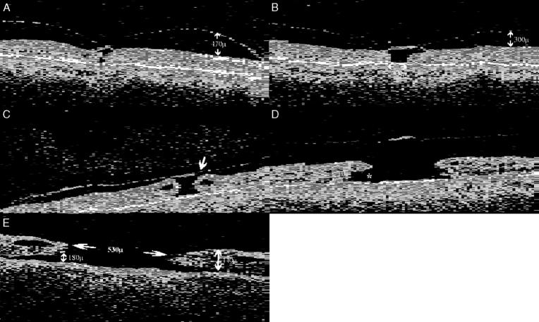
Figure 2 .
Evolution of idiopathic FTMHs on OCT. A 69 year old female with a long standing FTMH in the first eye and a 2 month history of central visual loss (acuity 6/12) and metamorphopsia in the fellow eye caused by a stage 1a lesion. OCT examination (A) revealed a central foveal detachment and a high reflectance interface in the preretinal plane which is thought to represent the posterior vitreous cortex, partially detached at the macula but remaining tethered to the central fovea. (B) Two months later a stage 1b lesion was noted on funduscopy and OCT revealed a more extensive foveal detachment. (C) At 4 months after the onset of symptoms, she had developed a stage 2 lesion with the acuity dropping to 6/24. The OCT confirms the presence of a pericentric full thickness break (arrow) and the early formation of an operculum. Cystic spaces are present at the edges of the hole. The patient elected to undergo surgical treatment at this stage. (D) An OCT of a stage 3 lesion in a 70 year old woman, showing a FTMH (380 µm) with vitreomacular separation and an operculum suspended on the posterior vitreous face. A prominent subretinal fluid cuff is visible (asterisk). (E) A stage 4 lesion in a 65 year old man. Note the extensive subretinal fluid cuff and the prominent cystoid spaces at the level of the inner plexiform layer at the edges of the hole.
Figure 3 .
Spontaneous arrest of a stage 1 lesion. (A) OCT of the fellow eye of a 64 year old woman with a stage 1a lesion on funduscopy and acuity of 6/9. The lesion was observed and 2 months later she reported an improvement in her metamorphopsia. Funduscopy revealed a reattached fovea with an inner lamellar defect. A free operculum was present in the preretinal plane with vitreomacular separation but otherwise attached vitreous. OCT (B) confirmed the clinical findings and no residual vitreofoveal traction. The visual acuity remained at 6/9 throughout follow up.
Figure 4 .
Spontaneous arrest and closure of a stage 2 lesion. (A) OCT of the first eye of a 56 year old female who presented with a stage 2 "can opener" lesion and a visual acuity of 6/36. Three months later she reported an improvement in the visual acuity (6/12) and funduscopy revealed a normal foveal reflex with vitreomacular separation and a free floating operculum. OCT (B) showed a relatively normal foveal contour, vitreomacular separation with an operculum, and no residual vitreofoveal traction.
Selected References
These references are in PubMed. This may not be the complete list of references from this article.
- Aaberg T. M., Blair C. J., Gass J. D. Macular holes. Am J Ophthalmol. 1970 Apr;69(4):555–562. doi: 10.1016/0002-9394(70)91620-x. [DOI] [PubMed] [Google Scholar]
- Akiba J., Yoshida A., Trempe C. L. Risk of developing a macular hole. Arch Ophthalmol. 1990 Aug;108(8):1088–1090. doi: 10.1001/archopht.1990.01070100044031. [DOI] [PubMed] [Google Scholar]
- Asrani S., Zeimer R., Goldberg M. F., Zou S. Serial optical sectioning of macular holes at different stages of development. Ophthalmology. 1998 Jan;105(1):66–77. doi: 10.1016/s0161-6420(98)91274-x. [DOI] [PubMed] [Google Scholar]
- Avila M. P., Jalkh A. E., Murakami K., Trempe C. L., Schepens C. L. Biomicroscopic study of the vitreous in macular breaks. Ophthalmology. 1983 Nov;90(11):1277–1283. doi: 10.1016/s0161-6420(83)34391-8. [DOI] [PubMed] [Google Scholar]
- Beausencourt E., Elsner A. E., Hartnett M. E., Trempe C. L. Quantitative analysis of macular holes with scanning laser tomography. Ophthalmology. 1997 Dec;104(12):2018–2029. doi: 10.1016/s0161-6420(97)30062-1. [DOI] [PubMed] [Google Scholar]
- Birch D. G., Jost B. F., Fish G. E. The focal electroretinogram in fellow eyes of patients with idiopathic macular holes. Arch Ophthalmol. 1988 Nov;106(11):1558–1563. doi: 10.1001/archopht.1988.01060140726043. [DOI] [PubMed] [Google Scholar]
- Bronstein M. A., Trempe C. L., Freeman H. M. Fellow eyes of eyes with macular holes. Am J Ophthalmol. 1981 Dec;92(6):757–761. doi: 10.1016/s0002-9394(14)75625-9. [DOI] [PubMed] [Google Scholar]
- CROLL L. J., CROLL M. Hole in the macula. Am J Ophthalmol. 1950 Feb;33(2):248-53, illust. doi: 10.1016/0002-9394(50)90844-0. [DOI] [PubMed] [Google Scholar]
- Campochiaro P. A., Van Niel E., Vinores S. A. Immunocytochemical labeling of cells in cortical vitreous from patients with premacular hole lesions. Arch Ophthalmol. 1992 Mar;110(3):371–377. doi: 10.1001/archopht.1992.01080150069031. [DOI] [PubMed] [Google Scholar]
- Chew E. Y., Sperduto R. D., Hiller R., Nowroozi L., Seigel D., Yanuzzi L. A., Burton T. C., Seddon J. M., Gragoudas E. S., Haller J. A. Clinical course of macular holes: the Eye Disease Case-Control Study. Arch Ophthalmol. 1999 Feb;117(2):242–246. doi: 10.1001/archopht.117.2.242. [DOI] [PubMed] [Google Scholar]
- Ezra E., Munro P. M., Charteris D. G., Aylward W. G., Luthert P. J., Gregor Z. J. Macular hole opercula. Ultrastructural features and clinicopathological correlation. Arch Ophthalmol. 1997 Nov;115(11):1381–1387. doi: 10.1001/archopht.1997.01100160551004. [DOI] [PubMed] [Google Scholar]
- Ezra E., Wells J. A., Gray R. H., Kinsella F. M., Orr G. M., Grego J., Arden G. B., Gregor Z. J. Incidence of idiopathic full-thickness macular holes in fellow eyes. A 5-year prospective natural history study. Ophthalmology. 1998 Feb;105(2):353–359. doi: 10.1016/s0161-6420(98)93562-x. [DOI] [PubMed] [Google Scholar]
- Fisher Y. L., Slakter J. S., Yannuzzi L. A., Guyer D. R. A prospective natural history study and kinetic ultrasound evaluation of idiopathic macular holes. Ophthalmology. 1994 Jan;101(1):5–11. doi: 10.1016/s0161-6420(94)31356-x. [DOI] [PubMed] [Google Scholar]
- Foos R. Y. Vitreoretinal juncture; topographical variations. Invest Ophthalmol. 1972 Oct;11(10):801–808. [PubMed] [Google Scholar]
- Freeman W. R., Azen S. P., Kim J. W., el-Haig W., Mishell D. R., 3rd, Bailey I. Vitrectomy for the treatment of full-thickness stage 3 or 4 macular holes. Results of a multicentered randomized clinical trial. The Vitrectomy for Treatment of Macular Hole Study Group. Arch Ophthalmol. 1997 Jan;115(1):11–21. doi: 10.1001/archopht.1997.01100150013002. [DOI] [PubMed] [Google Scholar]
- Funata M., Wendel R. T., de la Cruz Z., Green W. R. Clinicopathologic study of bilateral macular holes treated with pars plana vitrectomy and gas tamponade. Retina. 1992;12(4):289–298. doi: 10.1097/00006982-199212040-00001. [DOI] [PubMed] [Google Scholar]
- Gallemore R. P., Jumper J. M., McCuen B. W., 2nd, Jaffe G. J., Postel E. A., Toth C. A. Diagnosis of vitreoretinal adhesions in macular disease with optical coherence tomography. Retina. 2000;20(2):115–120. [PubMed] [Google Scholar]
- Gass J. D. Idiopathic senile macular hole. Its early stages and pathogenesis. Arch Ophthalmol. 1988 May;106(5):629–639. doi: 10.1001/archopht.1988.01060130683026. [DOI] [PubMed] [Google Scholar]
- Gass J. D., Joondeph B. C. Observations concerning patients with suspected impending macular holes. Am J Ophthalmol. 1990 Jun 15;109(6):638–646. doi: 10.1016/s0002-9394(14)72431-6. [DOI] [PubMed] [Google Scholar]
- Gass J. D. Müller cell cone, an overlooked part of the anatomy of the fovea centralis: hypotheses concerning its role in the pathogenesis of macular hole and foveomacualr retinoschisis. Arch Ophthalmol. 1999 Jun;117(6):821–823. doi: 10.1001/archopht.117.6.821. [DOI] [PubMed] [Google Scholar]
- Gass J. D. Reappraisal of biomicroscopic classification of stages of development of a macular hole. Am J Ophthalmol. 1995 Jun;119(6):752–759. doi: 10.1016/s0002-9394(14)72781-3. [DOI] [PubMed] [Google Scholar]
- Gaudric A., Haouchine B., Massin P., Paques M., Blain P., Erginay A. Macular hole formation: new data provided by optical coherence tomography. Arch Ophthalmol. 1999 Jun;117(6):744–751. doi: 10.1001/archopht.117.6.744. [DOI] [PubMed] [Google Scholar]
- Glaser B. M., Michels R. G., Kuppermann B. D., Sjaarda R. N., Pena R. A. Transforming growth factor-beta 2 for the treatment of full-thickness macular holes. A prospective randomized study. Ophthalmology. 1992 Jul;99(7):1162–1173. doi: 10.1016/s0161-6420(92)31837-8. [DOI] [PubMed] [Google Scholar]
- Guez J. E., Le Gargasson J. F., Massin P., Rigaudière F., Grall Y., Gaudric A. Functional assessment of macular hole surgery by scanning laser ophthalmoscopy. Ophthalmology. 1998 Apr;105(4):694–699. doi: 10.1016/S0161-6420(98)94026-X. [DOI] [PubMed] [Google Scholar]
- Guyer D. R., Green W. R., de Bustros S., Fine S. L. Histopathologic features of idiopathic macular holes and cysts. Ophthalmology. 1990 Aug;97(8):1045–1051. doi: 10.1016/s0161-6420(90)32465-x. [DOI] [PubMed] [Google Scholar]
- Guyer D. R., de Bustros S., Diener-West M., Fine S. L. Observations on patients with idiopathic macular holes and cysts. Arch Ophthalmol. 1992 Sep;110(9):1264–1268. doi: 10.1001/archopht.1992.01080210082030. [DOI] [PubMed] [Google Scholar]
- Hee M. R., Puliafito C. A., Wong C., Duker J. S., Reichel E., Schuman J. S., Swanson E. A., Fujimoto J. G. Optical coherence tomography of macular holes. Ophthalmology. 1995 May;102(5):748–756. doi: 10.1016/s0161-6420(95)30959-1. [DOI] [PubMed] [Google Scholar]
- Hikichi T., Yoshida A., Akiba J., Konno S., Trempe C. L. Prognosis of stage 2 macular holes. Am J Ophthalmol. 1995 May;119(5):571–575. doi: 10.1016/s0002-9394(14)70214-4. [DOI] [PubMed] [Google Scholar]
- Hudson C., Charles S. J., Flanagan J. G., Brahma A. K., Turner G. S., McLeod D. Objective morphological assessment of macular hole surgery by scanning laser tomography. Br J Ophthalmol. 1997 Feb;81(2):107–116. doi: 10.1136/bjo.81.2.107. [DOI] [PMC free article] [PubMed] [Google Scholar]
- James M., Feman S. S. Macular holes. Albrecht Von Graefes Arch Klin Exp Ophthalmol. 1980;215(1):59–63. doi: 10.1007/BF00413397. [DOI] [PubMed] [Google Scholar]
- Johnson R. N., Gass J. D. Idiopathic macular holes. Observations, stages of formation, and implications for surgical intervention. Ophthalmology. 1988 Jul;95(7):917–924. doi: 10.1016/s0161-6420(88)33075-7. [DOI] [PubMed] [Google Scholar]
- Jost B. F., Hutton W. L., Fuller D. G., Vaiser A., Snyder W. B., Fish G. E., Spencer R., Birch D. G. Vitrectomy in eyes at risk for macular hole formation. Ophthalmology. 1990 Jul;97(7):843–847. doi: 10.1016/s0161-6420(90)32493-4. [DOI] [PubMed] [Google Scholar]
- Kakehashi A., Schepens C. L., Akiba J., Hikichi T., Trempe C. L. Spontaneous resolution of foveal detachments and macular breaks. Am J Ophthalmol. 1995 Dec;120(6):767–775. doi: 10.1016/s0002-9394(14)72730-8. [DOI] [PubMed] [Google Scholar]
- Kalina R. E., Wells C. G. Screening for ocular toxicity in asymptomatic patients treated with tamoxifen. Am J Ophthalmol. 1995 Jan;119(1):112–113. doi: 10.1016/s0002-9394(14)73835-8. [DOI] [PubMed] [Google Scholar]
- Kelly N. E., Wendel R. T. Vitreous surgery for idiopathic macular holes. Results of a pilot study. Arch Ophthalmol. 1991 May;109(5):654–659. doi: 10.1001/archopht.1991.01080050068031. [DOI] [PubMed] [Google Scholar]
- Kim J. W., Freeman W. R., Azen S. P., el-Haig W., Klein D. J., Bailey I. L. Prospective randomized trial of vitrectomy or observation for stage 2 macular holes. Vitrectomy for Macular Hole Study Group. Am J Ophthalmol. 1996 Jun;121(6):605–614. doi: 10.1016/s0002-9394(14)70625-7. [DOI] [PubMed] [Google Scholar]
- Kim J. W., Freeman W. R., el-Haig W., Maguire A. M., Arevalo J. F., Azen S. P. Baseline characteristics, natural history, and risk factors to progression in eyes with stage 2 macular holes. Results from a prospective randomized clinical trial. Vitrectomy for Macular Hole Study Group. Ophthalmology. 1995 Dec;102(12):1818–1829. doi: 10.1016/s0161-6420(95)30788-9. [DOI] [PubMed] [Google Scholar]
- Kishi S., Demaria C., Shimizu K. Vitreous cortex remnants at the fovea after spontaneous vitreous detachment. Int Ophthalmol. 1986 Dec;9(4):253–260. doi: 10.1007/BF00137539. [DOI] [PubMed] [Google Scholar]
- Kishi S., Shimizu K. Posterior precortical vitreous pocket. Arch Ophthalmol. 1990 Jul;108(7):979–982. doi: 10.1001/archopht.1990.01070090081044. [DOI] [PubMed] [Google Scholar]
- Lansing M. B., Glaser B. M., Liss H., Hanham A., Thompson J. T., Sjaarda R. N., Gordon A. J. The effect of pars plana vitrectomy and transforming growth factor-beta 2 without epiretinal membrane peeling on full-thickness macular holes. Ophthalmology. 1993 Jun;100(6):868–872. doi: 10.1016/s0161-6420(93)31561-7. [DOI] [PubMed] [Google Scholar]
- Lewis M. L., Cohen S. M., Smiddy W. E., Gass J. D. Bilaterality of idiopathic macular holes. Graefes Arch Clin Exp Ophthalmol. 1996 Apr;234(4):241–245. doi: 10.1007/BF00430416. [DOI] [PubMed] [Google Scholar]
- Madreperla S. A., Geiger G. L., Funata M., de la Cruz Z., Green W. R. Clinicopathologic correlation of a macular hole treated by cortical vitreous peeling and gas tamponade. Ophthalmology. 1994 Apr;101(4):682–686. doi: 10.1016/s0161-6420(94)31278-4. [DOI] [PubMed] [Google Scholar]
- Madreperla S. A., McCuen B. W., 2nd, Hickingbotham D., Green W. R. Clinicopathologic correlation of surgically removed macular hole opercula. Am J Ophthalmol. 1995 Aug;120(2):197–207. doi: 10.1016/s0002-9394(14)72608-x. [DOI] [PubMed] [Google Scholar]
- Margheria R. R., Schepens C. L. Macular breaks. 1. Diagnosis, etiology, and observations. Am J Ophthalmol. 1972 Aug;74(2):219–232. [PubMed] [Google Scholar]
- Margherio R. R., Trese M. T., Margherio A. R., Cartright K. Surgical management of vitreomacular traction syndromes. Ophthalmology. 1989 Sep;96(9):1437–1445. doi: 10.1016/s0161-6420(89)32711-4. [DOI] [PubMed] [Google Scholar]
- McDonnell P. J., Fine S. L., Hillis A. I. Clinical features of idiopathic macular cysts and holes. Am J Ophthalmol. 1982 Jun;93(6):777–786. doi: 10.1016/0002-9394(82)90474-3. [DOI] [PubMed] [Google Scholar]
- Morgan C. M., Schatz H. Idiopathic macular holes. Am J Ophthalmol. 1985 Apr 15;99(4):437–444. doi: 10.1016/0002-9394(85)90011-x. [DOI] [PubMed] [Google Scholar]
- Morgan C. M., Schatz H. Involutional macular thinning. A pre-macular hole condition. Ophthalmology. 1986 Feb;93(2):153–161. doi: 10.1016/s0161-6420(86)33767-9. [DOI] [PubMed] [Google Scholar]
- Mori K., Abe T., Yoneya S. Dome-shaped detachment of premacular vitreous cortex in macular hole development. Ophthalmic Surg Lasers. 2000 May-Jun;31(3):203–209. [PubMed] [Google Scholar]
- Noyes H. D. Detachment of Retina with Laceration at Macula. Trans Am Ophthalmol Soc. 1871;1(8):128–129. [PMC free article] [PubMed] [Google Scholar]
- Orellana J., Lieberman R. M. Stage III macular hole surgery. Br J Ophthalmol. 1993 Sep;77(9):555–558. doi: 10.1136/bjo.77.9.555. [DOI] [PMC free article] [PubMed] [Google Scholar]
- Rosa R. H., Jr, Glaser B. M., de la Cruz Z., Green W. R. Clinicopathologic correlation of an untreated macular hole and a macular hole treated by vitrectomy, transforming growth factor-beta 2, and gas tamponade. Am J Ophthalmol. 1996 Dec;122(6):853–863. doi: 10.1016/s0002-9394(14)70382-4. [DOI] [PubMed] [Google Scholar]
- Roth A. M., Foos R. Y. Surface wrinkling retinopathy in eyes enucleated at autopsy. Trans Am Acad Ophthalmol Otolaryngol. 1971 Sep-Oct;75(5):1047–1058. [PubMed] [Google Scholar]
- Ruby A. J., Williams D. F., Grand M. G., Thomas M. A., Meredith T. A., Boniuk I., Olk R. J. Pars plana vitrectomy for treatment of stage 2 macular holes. Arch Ophthalmol. 1994 Mar;112(3):359–364. doi: 10.1001/archopht.1994.01090150089029. [DOI] [PubMed] [Google Scholar]
- Ryan E. H., Jr, Gilbert H. D. Results of surgical treatment of recent-onset full-thickness idiopathic macular holes. Arch Ophthalmol. 1994 Dec;112(12):1545–1553. doi: 10.1001/archopht.1994.01090240051025. [DOI] [PubMed] [Google Scholar]
- Smiddy W. E., Gass J. D. Masquerades of macular holes. Ophthalmic Surg. 1995 Jan-Feb;26(1):16–24. [PubMed] [Google Scholar]
- Smiddy W. E., Glaser B. M., Thompson J. T., Sjaarda R. N., Flynn H. W., Jr, Hanham A., Murphy R. P. Transforming growth factor-beta 2 significantly enhances the ability to flatten the rim of subretinal fluid surrounding macular holes. Preliminary anatomic results of a multicenter prospective randomized study. Retina. 1993;13(4):296–301. [PubMed] [Google Scholar]
- Smiddy W. E., Michels R. G., Glaser B. M., de Bustros S. Vitrectomy for impending idiopathic macular holes. Am J Ophthalmol. 1988 Apr 15;105(4):371–376. doi: 10.1016/0002-9394(88)90300-5. [DOI] [PubMed] [Google Scholar]
- Smiddy W. E., Michels R. G., de Bustros S., de la Cruz Z., Green W. R. Histopathology of tissue removed during vitrectomy for impending idiopathic macular holes. Am J Ophthalmol. 1989 Oct 15;108(4):360–364. doi: 10.1016/s0002-9394(14)73301-x. [DOI] [PubMed] [Google Scholar]
- Trempe C. L., Weiter J. J., Furukawa H. Fellow eyes in cases of macular hole. Biomicroscopic study of the vitreous. Arch Ophthalmol. 1986 Jan;104(1):93–95. doi: 10.1001/archopht.1986.01050130103031. [DOI] [PubMed] [Google Scholar]
- Watzke R. C., Allen L. Subjective slitbeam sign for macular disease. Am J Ophthalmol. 1969 Sep;68(3):449–453. doi: 10.1016/0002-9394(69)90712-0. [DOI] [PubMed] [Google Scholar]
- Weinberger D., Stiebel H., Gaton D. D., Priel E., Yassur Y. Three-dimensional measurements of idiopathic macular holes using a scanning laser tomograph. Ophthalmology. 1995 Oct;102(10):1445–1449. doi: 10.1016/s0161-6420(95)30847-0. [DOI] [PubMed] [Google Scholar]
- Wells J. A., Gregor Z. J. Surgical treatment of full-thickness macular holes using autologous serum. Eye (Lond) 1996;10(Pt 5):593–599. doi: 10.1038/eye.1996.136. [DOI] [PubMed] [Google Scholar]
- Wendel R. T., Patel A. C., Kelly N. E., Salzano T. C., Wells J. W., Novack G. D. Vitreous surgery for macular holes. Ophthalmology. 1993 Nov;100(11):1671–1676. doi: 10.1016/s0161-6420(93)31419-3. [DOI] [PubMed] [Google Scholar]
- Yaoeda H. [Clinical observation on the macular hole]. Nippon Ganka Gakkai Zasshi. 1967 Sep;71(9):1723–1736. [PubMed] [Google Scholar]
- de Bustros S. Vitrectomy for prevention of macular hole study. Arch Ophthalmol. 1991 Aug;109(8):1057–1057. doi: 10.1001/archopht.1991.01080080015003. [DOI] [PubMed] [Google Scholar]
- de Bustros S. Vitrectomy for prevention of macular holes. Results of a randomized multicenter clinical trial. Vitrectomy for Prevention of Macular Hole Study Group. Ophthalmology. 1994 Jun;101(6):1055–1060. doi: 10.1016/s0161-6420(94)31218-8. [DOI] [PubMed] [Google Scholar]
- von Rückmann A., Fitzke F. W., Gregor Z. J. Fundus autofluorescence in patients with macular holes imaged with a laser scanning ophthalmoscope. Br J Ophthalmol. 1998 Apr;82(4):346–351. doi: 10.1136/bjo.82.4.346. [DOI] [PMC free article] [PubMed] [Google Scholar]


