Full Text
The Full Text of this article is available as a PDF (325.9 KB).
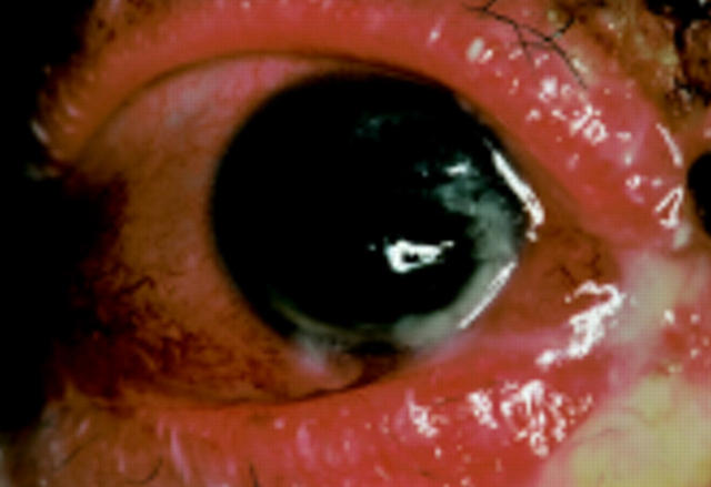
Figure 1 .
Photograph of the anterior segment of the right eye on 13 April 1999.
Figure 2 .
Histological examination by haematoxylin and eosin staining of lesional skin disclosed intraepidermal clefts which contained several acantholytic cells (left). The direct immunofluorescent staining of the skin showed intercellular deposition of immunogloblin G (right).


