Abstract
AIMS—To clarify the developmental mechanism and critical period for the uncommon complex of Peters' anomaly and persistent hyperplastic primary vitreous (PHPV). METHODS—Two eyes with Peters' anomaly and PHPV were histologically examined by serial section. One eye was enucleated at age 7 months (case 1) and the other at age 4 months (case 2) owing to severe anterior staphyloma. RESULTS—In both eyes, defects in the endothelium, Descemet's membrane, and posterior stroma were observed in the central cornea, and the degenerative lens adhered to the posterior surface of the defective corneal stroma. Also, in both eyes, the anterior chamber space was not formed and the undifferentiated iris stroma adhered to the posterior surface of the peripheral cornea. Mesenchymal tissue containing melanocytes was observed behind the degenerative lens, and the pigment epithelium was absent at the lower nasal side of the ciliary body in case 1. In case 2, mesenchymal tissue containing scattered melanocytes in the vitreous cavity was seen on the posterior retina. Based on the histological findings, both cases were diagnosed as Peters' anomaly caused by the faulty separation of the lens vesicle, PHPV, maldevelopment of the iris and ciliary body, and goniodysgenesis. CONCLUSION—Migratory disorders of neural crest cells from 4 to 7 weeks of gestation may be responsible for various ocular anomalies including Peters' anomaly and PHPV, as observed in these cases.
Full Text
The Full Text of this article is available as a PDF (152.4 KB).
Figure 1 .
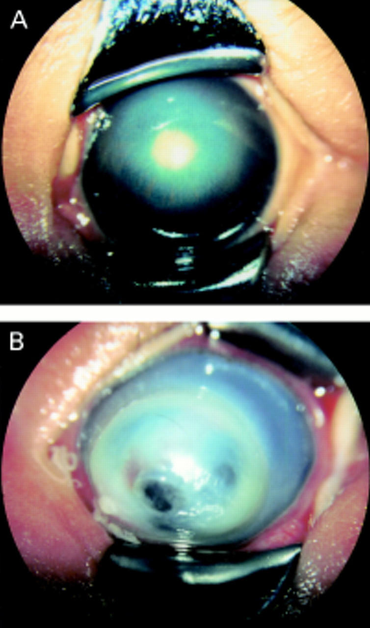
(A) A photograph of anterior segment of case 1. Central corneal opacity, elongated ciliary processes, and whitish mass behind the lens are observed. (B) A photograph of anterior segment of case 2. The cornea is noticeably protruding and densely opaque overall. These findings correspond to anterior staphyloma.
Figure 2 .
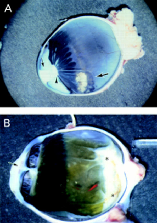
(A) Photograph of the section of the eyeball of case 1. The degenerative lens (small arrow) adheres to the defective central cornea, the whitish mass (arrowhead) is observed behind the lens, and ciliary coloboma is seen (large arrow). Elongated ciliary processes are also observed. (B) Photograph of the section of the eyeball of case 2. The degenerative lens (arrow) adheres to the defective central cornea, and the whitish membranous tissue (asterisks) is observed on the posterior retina.
Figure 3 .
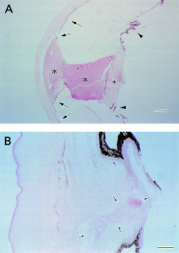
(A). A histological photograph of the anterior segment of case 1. The degenerative lens (large asterisks) adheres to the posterior surface of the defective corneal stroma. The anterior chamber space is not formed, and the undifferentiated iris stroma (arrows) adheres to the posterior surface of the peripheral cornea and lens. Mesenchymal tissue (small asterisk) is observed behind the degenerative lens. Elongated ciliary processes (arrowheads) are also observed. Haematoxylin and eosin. Bar = 700 µm. (B). A histological photograph of the anterior segment of case 2. In the central cornea, the defects in the endothelium, Descemet's membrane, and posterior stroma are observed, and the lens material (arrows) is detected in the defective corneal stroma. The anterior chamber space is not formed, and the undifferentiated iris stroma (asterisks) adheres to the posterior surface of the peripheral cornea. Haematoxylin and eosin. Bar = 280 µm.
Figure 4 .
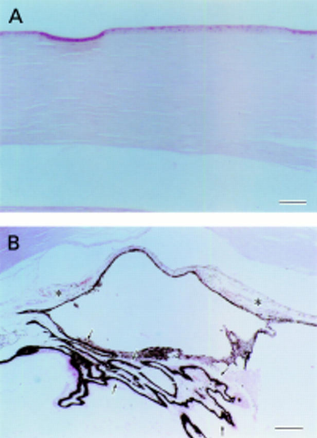
(A) A histological photograph of the central cornea of case 1. In the central cornea, defects in the endothelium, Descemet's membrane, and posterior stroma are observed. Haematoxylin and eosin. Bar = 70 µm. (B) A histological photograph of the iris, ciliary body, and trabecular meshwork of case 2. The iris (asterisks), ciliary body (arrows), and trabecular meshwork are poorly differentiated, and Schlemm's canal is not observed. Haematoxylin and eosin. Bar = 175 µm.
Figure 5 .
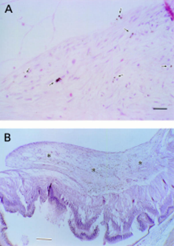
(A) A histological photograph of the mesenchymal tissue behind the lens of case 1. A few melanocytes (arrows) are observed in the mesenchymal tissue. Haematoxylin and eosin. Bar = 28 µm. (B) A histological photograph of the mesenchymal tissue on the retina of case 2. Mesenchymal tissue (asterisks) containing scattered melanocytes in the vitreous cavity are seen on the retina, inducing folds in the neural retina. Haematoxylin and eosin. Bar = 280 µm.
Selected References
These references are in PubMed. This may not be the complete list of references from this article.
- Bahn C. F., Falls H. F., Varley G. A., Meyer R. F., Edelhauser H. F., Bourne W. M. Classification of corneal endothelial disorders based on neural crest origin. Ophthalmology. 1984 Jun;91(6):558–563. doi: 10.1016/s0161-6420(84)34249-x. [DOI] [PubMed] [Google Scholar]
- Beauchamp G. R., Knepper P. A. Role of the neural crest in anterior segment development and disease. J Pediatr Ophthalmol Strabismus. 1984 Nov-Dec;21(6):209–214. doi: 10.3928/0191-3913-19841101-03. [DOI] [PubMed] [Google Scholar]
- Cook C. S. Experimental models of anterior segment dysgenesis. Ophthalmic Paediatr Genet. 1989 Mar;10(1):33–46. doi: 10.3109/13816818909083771. [DOI] [PubMed] [Google Scholar]
- Cook C. S., Sulik K. K. Keratolenticular dysgenesis (Peters' anomaly) as a result of acute embryonic insult during gastrulation. J Pediatr Ophthalmol Strabismus. 1988 Mar-Apr;25(2):60–66. doi: 10.3928/0191-3913-19880301-04. [DOI] [PubMed] [Google Scholar]
- Haddad R., Font R. L., Reeser F. Persistent hyperplastic primary vitreous. A clinicopathologic study of 62 cases and review of the literature. Surv Ophthalmol. 1978 Sep-Oct;23(2):123–134. doi: 10.1016/0039-6257(78)90091-7. [DOI] [PubMed] [Google Scholar]
- Heon E., Barsoum-Homsy M., Cevrette L., Jacob J. L., Milot J., Polemeno R., Musarella M. A. Peters' anomaly. The spectrum of associated ocular and systemic malformations. Ophthalmic Paediatr Genet. 1992 Jun;13(2):137–143. doi: 10.3109/13816819209087614. [DOI] [PubMed] [Google Scholar]
- Johnston M. C., Noden D. M., Hazelton R. D., Coulombre J. L., Coulombre A. J. Origins of avian ocular and periocular tissues. Exp Eye Res. 1979 Jul;29(1):27–43. doi: 10.1016/0014-4835(79)90164-7. [DOI] [PubMed] [Google Scholar]
- Mann I. CONGENITAL RETINAL FOLD. Br J Ophthalmol. 1935 Dec;19(12):641–658. doi: 10.1136/bjo.19.12.641. [DOI] [PMC free article] [PubMed] [Google Scholar]
- Mayer U. M. Peters' anomaly and combination with other malformations (series of 16 patients). Ophthalmic Paediatr Genet. 1992 Jun;13(2):131–135. doi: 10.3109/13816819209087613. [DOI] [PubMed] [Google Scholar]
- Myles W. M., Flanders M. E., Chitayat D., Brownstein S. Peters' anomaly: a clinicopathologic study. J Pediatr Ophthalmol Strabismus. 1992 Nov-Dec;29(6):374–381. doi: 10.3928/0191-3913-19921101-10. [DOI] [PubMed] [Google Scholar]
- Ozeki H., Shirai S. Developmental eye abnormalities in mouse fetuses induced by retinoic acid. Jpn J Ophthalmol. 1998 May-Jun;42(3):162–167. doi: 10.1016/s0021-5155(97)00134-2. [DOI] [PubMed] [Google Scholar]
- Ozeki H., Shirai S., Ikeda K., Ogura Y. Critical period for retinoic acid-induced developmental abnormalities of the vitreous in mouse fetuses. Exp Eye Res. 1999 Feb;68(2):223–228. doi: 10.1006/exer.1998.0591. [DOI] [PubMed] [Google Scholar]
- Ozeki H., Shirai S., Nozaki M., Ikeda K., Ogura Y. Maldevelopment of neural crest cells in patients with typical uveal coloboma. J Pediatr Ophthalmol Strabismus. 1999 Nov-Dec;36(6):337–341. doi: 10.3928/0191-3913-19991101-09. [DOI] [PubMed] [Google Scholar]
- REESE A. B. Persistent hyperplastic primary vitreous. Am J Ophthalmol. 1955 Sep;40(3):317–331. doi: 10.1016/0002-9394(55)91866-3. [DOI] [PubMed] [Google Scholar]
- Shirai S. [Developmental mechanisms of congenital eye abnormalities]. Nippon Ganka Gakkai Zasshi. 1991 Dec;95(12):1206–1237. [PubMed] [Google Scholar]
- Uemura Y. [Clinico-anatomical studies on the developing vitreous and retina]. Nippon Ganka Gakkai Zasshi. 1986 Jan;90(1):1–24. [PubMed] [Google Scholar]
- van Schooneveld M. J., Delleman J. W., Beemer F. A., Bleeker-Wagemakers E. M. Peters'-plus: a new syndrome. Ophthalmic Paediatr Genet. 1984 Dec;4(3):141–145. doi: 10.3109/13816818409006113. [DOI] [PubMed] [Google Scholar]


