Abstract
AIMS—To report the occurrence of paracentral scotomata after pars plana vitrectomy for idiopathic macular holes. METHODS—In 15 patients static microperimetry using a Rodenstock scanning laser ophthalmoscope (SLO-105) was performed preoperatively and 6 or 12 weeks postoperatively (stimulus size 0.2° (Goldmann II), employed intensity 0 and 12 dB, 20° fields in all tests). Surgery consisted of standard three port vitrectomy including removal of epiretinal membranes and the inner limiting membrane. RESULTS—Postoperative paracentral scotomata were detected in areas that were tested normally before surgery. They were mostly located temporally and/or inferiorly and often appeared like nerve fibre bundle defects. The greatest dimension varied from 1.2° to 4.0° (360-1200 µm), smallest dimension from 0.25° to 2.0° (75-600 µm). In three patients more than one scotoma was observed. CONCLUSION—Small, mostly asymptomatic, paracentral scotomata as a complication after vitrectomy for idiopathic macular hole have not been reported in the literature so far. Whether they are caused by trauma to the nerve fibres during surgery or other factors remains unknown.
Full Text
The Full Text of this article is available as a PDF (151.8 KB).
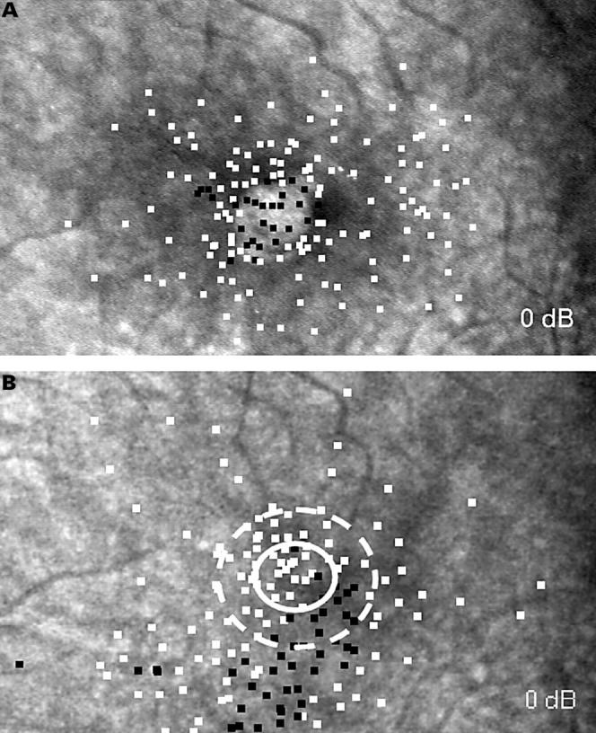
Figure 1 .

(A) Preoperative and postoperative (B) microperimetry of a left eye. White squares represent stimuli that were seen by the patient, black squares represent relative (12 dB) or deep (0 dB) scotomata. The white circle marks the area of the preoperative macular defect, the broken circle the area of the preoperative neurosensory detachment. Postoperatively a large deep scotoma inferior to the closed macular hole can be seen in an area that was tested normally before surgery. The scotoma had the shape of a nerve fibre bundle defect.
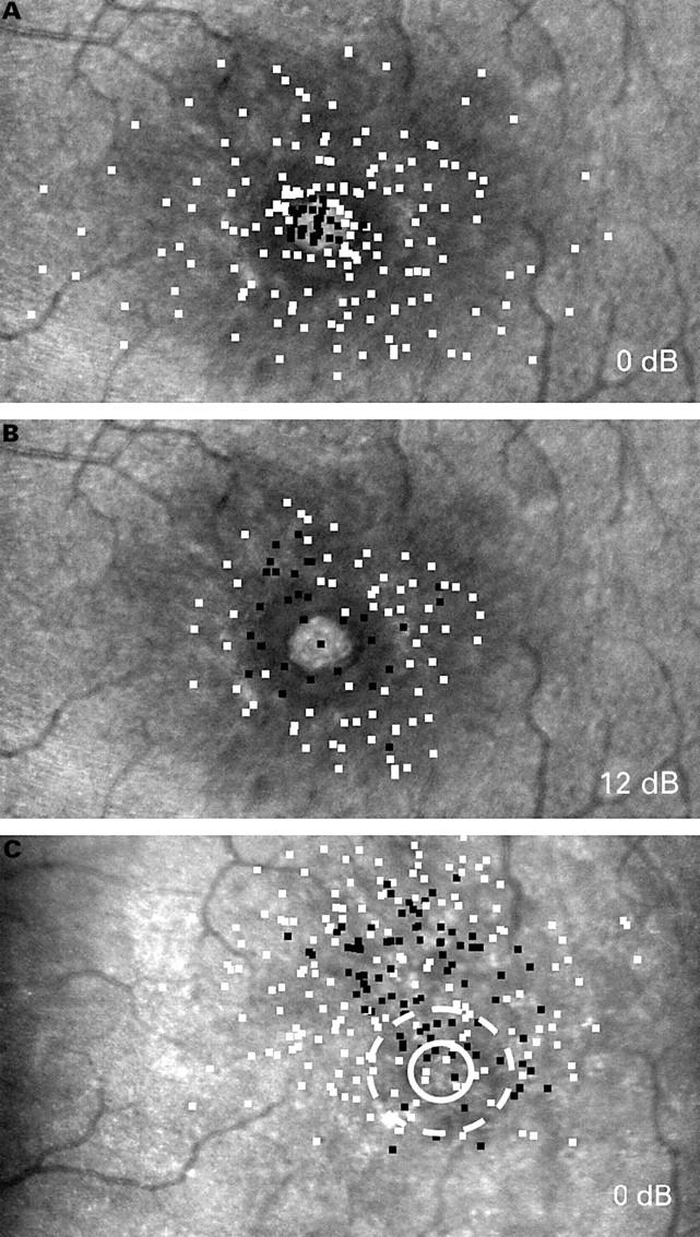
Figure 2 .

(A, B) Preoperative and postoperative (C) microperimetry of a left eye. Preoperatively a deep scotoma in the macular hole area is seen (A),which is surrounded by a relative scotoma (B). Postoperatively a large deep scotoma with irregular boundaries was found temporal to the area of the closed macular hole (C).
Selected References
These references are in PubMed. This may not be the complete list of references from this article.
- Acosta F., Lashkari K., Reynaud X., Jalkh A. E., Van de Velde F., Chedid N. Characterization of functional changes in macular holes and cysts. Ophthalmology. 1991 Dec;98(12):1820–1823. doi: 10.1016/s0161-6420(91)32044-x. [DOI] [PubMed] [Google Scholar]
- Boldt H. C., Munden P. M., Folk J. C., Mehaffey M. G. Visual field defects after macular hole surgery. Am J Ophthalmol. 1996 Sep;122(3):371–381. doi: 10.1016/s0002-9394(14)72064-1. [DOI] [PubMed] [Google Scholar]
- Byhr E., Lindblom B. Preoperative measurements of macular hole with scanning laser ophthalmoscopy. Correlation with functional outcome. Acta Ophthalmol Scand. 1998 Oct;76(5):579–583. doi: 10.1034/j.1600-0420.1998.760513.x. [DOI] [PubMed] [Google Scholar]
- Freeman W. R., Azen S. P., Kim J. W., el-Haig W., Mishell D. R., 3rd, Bailey I. Vitrectomy for the treatment of full-thickness stage 3 or 4 macular holes. Results of a multicentered randomized clinical trial. The Vitrectomy for Treatment of Macular Hole Study Group. Arch Ophthalmol. 1997 Jan;115(1):11–21. doi: 10.1001/archopht.1997.01100150013002. [DOI] [PubMed] [Google Scholar]
- Freeman W. R. Vitrectomy surgery for full-thickness macular holes. Am J Ophthalmol. 1993 Aug 15;116(2):233–235. doi: 10.1016/s0002-9394(14)71292-9. [DOI] [PubMed] [Google Scholar]
- Gass J. D. Idiopathic senile macular hole. Its early stages and pathogenesis. Arch Ophthalmol. 1988 May;106(5):629–639. doi: 10.1001/archopht.1988.01060130683026. [DOI] [PubMed] [Google Scholar]
- Guez J. E., Le Gargasson J. F., Massin P., Rigaudière F., Grall Y., Gaudric A. Functional assessment of macular hole surgery by scanning laser ophthalmoscopy. Ophthalmology. 1998 Apr;105(4):694–699. doi: 10.1016/S0161-6420(98)94026-X. [DOI] [PubMed] [Google Scholar]
- Kelly N. E., Wendel R. T. Vitreous surgery for idiopathic macular holes. Results of a pilot study. Arch Ophthalmol. 1991 May;109(5):654–659. doi: 10.1001/archopht.1991.01080050068031. [DOI] [PubMed] [Google Scholar]
- Llobet A., Gual A., Palés J., Barraquer R., Tobías E., Nicolás J. M. Bradykinin decreases outflow facility in perfused anterior segments and induces shape changes in passaged BTM cells in vitro. Invest Ophthalmol Vis Sci. 1999 Jan;40(1):113–125. [PubMed] [Google Scholar]
- Melberg N. S., Thomas M. A. Visual field loss after pars plana vitrectomy with air/fluid exchange. Am J Ophthalmol. 1995 Sep;120(3):386–388. doi: 10.1016/s0002-9394(14)72169-5. [DOI] [PubMed] [Google Scholar]
- Park S. S., Marcus D. M., Duker J. S., Pesavento R. D., Topping T. M., Frederick A. R., Jr, D'Amico D. J. Posterior segment complications after vitrectomy for macular hole. Ophthalmology. 1995 May;102(5):775–781. doi: 10.1016/s0161-6420(95)30956-6. [DOI] [PubMed] [Google Scholar]
- Ruby A. J., Williams D. F., Grand M. G., Thomas M. A., Meredith T. A., Boniuk I., Olk R. J. Pars plana vitrectomy for treatment of stage 2 macular holes. Arch Ophthalmol. 1994 Mar;112(3):359–364. doi: 10.1001/archopht.1994.01090150089029. [DOI] [PubMed] [Google Scholar]
- Sjaarda R. N., Frank D. A., Glaser B. M., Thompson J. T., Murphy R. P. Assessment of vision in idiopathic macular holes with macular microperimetry using the scanning laser ophthalmoscope. Ophthalmology. 1993 Oct;100(10):1513–1518. doi: 10.1016/s0161-6420(93)31448-x. [DOI] [PubMed] [Google Scholar]
- Sjaarda R. N., Frank D. A., Glaser B. M., Thompson J. T., Murphy R. P. Resolution of an absolute scotoma and improvement of relative scotomata after successful macular hole surgery. Am J Ophthalmol. 1993 Aug 15;116(2):129–139. doi: 10.1016/s0002-9394(14)71276-0. [DOI] [PubMed] [Google Scholar]
- Wendel R. T., Patel A. C., Kelly N. E., Salzano T. C., Wells J. W., Novack G. D. Vitreous surgery for macular holes. Ophthalmology. 1993 Nov;100(11):1671–1676. doi: 10.1016/s0161-6420(93)31419-3. [DOI] [PubMed] [Google Scholar]


