Abstract
BACKGROUND—Anterior segment imaging using fluorescein angiography is only suitable in lightly pigmented irides as the brown pigmentation of the iris masks fluorescein transmission. Indocyanine green (ICG) angiography has excellent penetration of pigment epithelium and, therefore, has potential application in detecting perfusion changes of dark irides after strabismus surgery. METHODS—A prospective study was conducted on patients older than 15 years undergoing strabismus surgery. A fundus camera was focused on the arteriolar tufts of the pupillary margin and 50 mg of ICG (concentration of 12.5 mg/ml) was given intravenously. Images were then obtained at 1 minute intervals of 5 minutes' duration. RESULTS—45 patients with a mean age of 54.6 years and a mean follow up period of 8.6 weeks were studied. There were 23 patients in the primary surgery group, 11 in the secondary surgery group, and 11 in the staged group. Iris ICG angiograms were successfully performed in all patients. No persistent filling defect was detected in the primary and secondary horizontal recti surgery groups or in the secondary or staged vertical and combined vertical rectus groups 6-8 weeks postoperatively. 57% of both primary vertical and combined vertical and horizontal groups showed defects in the early postoperative phase. Only three cases demonstrated late perfusion defects in this series. CONCLUSION—ICG can detect iris perfusion changes in dark irides after strabismus surgery. Iris reperfusion was achieved in the majority of the cases.
Full Text
The Full Text of this article is available as a PDF (152.0 KB).
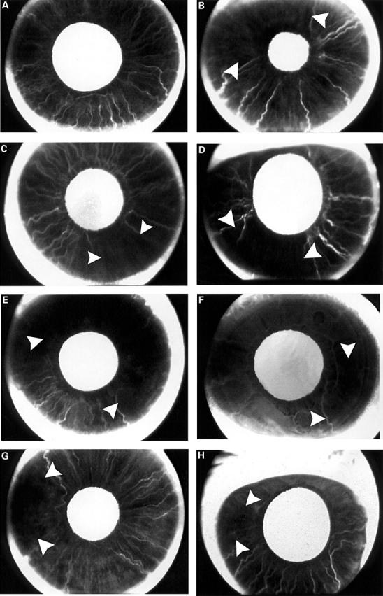
Figure 1 .

(A) Normal dark brown iris ICGA of the right eye in a 49 year old man. Late frame at 46 seconds showed complete filling of the iris. (B) ICGA of the right eye in a 68 year old man. One day after primary SR surgery. Superior and temporal sectors (arrowheads) filled out of phase by 34 seconds. (C) ICGA of the left eye in a 52 year old man. One day after primary IR surgery. The nasal, superior, and temporal sectors started to fill first which was followed by a delay in filling in inferior quadrant (arrowheads) by 40.2 seconds. (D) ICGA of the right eye in a 55 year old woman. One day after primary SR and IR surgery. Superior and inferior temporal sectors (arrowheads) showed delay in perfusion by 25 seconds. (E) ICGA of the left eye in a 59 year old man. One day after primary SR and MR surgery. Persistent superior sector delay (arrowheads) by 6 minutes; no nasal sector delay in filling. (F) ICGA of the left eye (previous 5 month history of LR and MR surgery) in a 58 year old woman. One day after IR and LR surgery. Inferior and temporal sectors filled out of phase (arrowheads). Nasal sector perfusion was normal. (G) ICGA of the right eye (previous 3 year history of MR surgery) in a 81 year old man. Preoperative ICGA showed persistent temporal sector delay at late frames. Change of surgical plan and limited surgery to LR only in this eye. One day after LR surgery. Temporal sector delay (arrowheads) persisted at 38.8 seconds. (H) ICGA of the right eye (previous 18 month history of SR and IR surgery) in a 15 year old girl. One day after MR and LR surgery. Temporal sector delay (arrowheads) in filling of 27.9 seconds and nasal sector was well perfused at early frames because of the MR anterior ciliary artery preservation technique 2 months after surgery.
Selected References
These references are in PubMed. This may not be the complete list of references from this article.
- CHERRICK G. R., STEIN S. W., LEEVY C. M., DAVIDSON C. S. Indocyanine green: observations on its physical properties, plasma decay, and hepatic extraction. J Clin Invest. 1960 Apr;39:592–600. doi: 10.1172/JCI104072. [DOI] [PMC free article] [PubMed] [Google Scholar]
- Hayreh S. S., Scott W. E. Fluorescein iris angiography. I. Normal pattern. Arch Ophthalmol. 1978 Aug;96(8):1383–1389. doi: 10.1001/archopht.1978.03910060137009. [DOI] [PubMed] [Google Scholar]
- Hayreh S. S., Scott W. E. Fluorescein iris angiography. II. Disturbances in iris circulation following strabismus operation on the various recti. Arch Ophthalmol. 1978 Aug;96(8):1390–1400. doi: 10.1001/archopht.1978.03910060144010. [DOI] [PubMed] [Google Scholar]
- Olver J. M., Lee J. P. Recovery of anterior segment circulation after strabismus surgery in adult patients. Ophthalmology. 1992 Mar;99(3):305–315. doi: 10.1016/s0161-6420(92)31971-2. [DOI] [PubMed] [Google Scholar]
- Olver J. M., Lee J. P. The effects of strabismus surgery on anterior segment circulation. Eye (Lond) 1989;3(Pt 3):318–326. doi: 10.1038/eye.1989.46. [DOI] [PubMed] [Google Scholar]
- Saunders R. A., Bluestein E. C., Wilson M. E., Berland J. E. Anterior segment ischemia after strabismus surgery. Surv Ophthalmol. 1994 Mar-Apr;38(5):456–466. doi: 10.1016/0039-6257(94)90175-9. [DOI] [PubMed] [Google Scholar]
- Saunders R. A., Phillips M. S. Anterior segment ischemia after three rectus muscle surgery. Ophthalmology. 1988 Apr;95(4):533–537. doi: 10.1016/s0161-6420(88)33154-4. [DOI] [PubMed] [Google Scholar]
- Yannuzzi L. A., Slakter J. S., Sorenson J. A., Guyer D. R., Orlock D. A. Digital indocyanine green videoangiography and choroidal neovascularization. Retina. 1992;12(3):191–223. [PubMed] [Google Scholar]


