Full Text
The Full Text of this article is available as a PDF (431.6 KB).
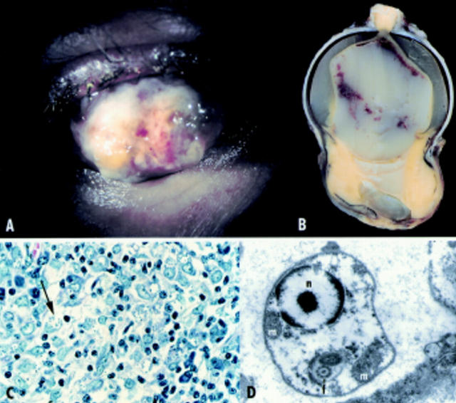
Figure 1 .
(A) Clinical examination of the right eye. Severe scleromalacia with focal perforation. (B) Macroscopical examination. Perforated sclera, lens, parts of the iris, and vitreous body luxated into the bulge. (C) Light microscopic examination. Leishmaniae (arrow) within histiocytes by Giemsa staining. (D) Transmission electron microscopic examination The protozoon consists of a nucleus (n), prominent mitochondria (m), and the flagellum (f).


