

Full Text
The Full Text of this article is available as a PDF (172.7 KB).
Figure 1 .
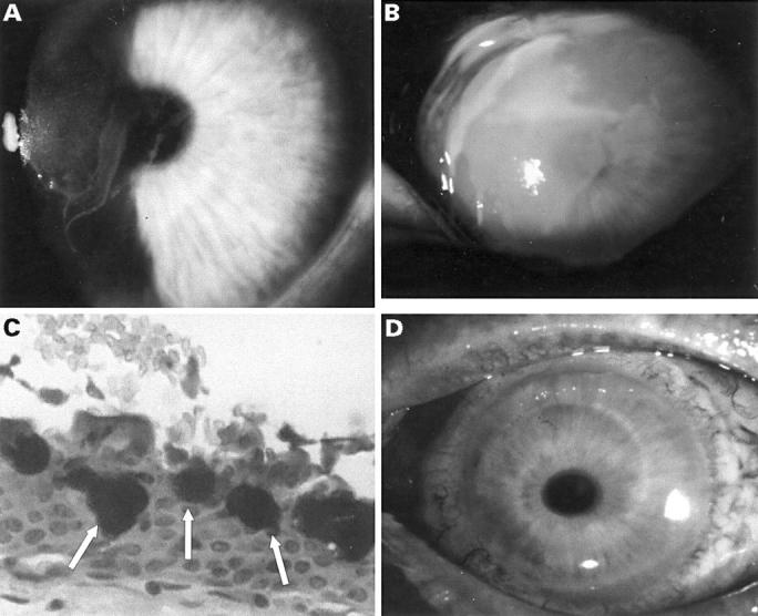
(A) Severe persistent epithelial defect on the right cornea after 2 years of oral hydroxyurea treatment. (B) Eighteen days after reintroduction of hydroxyurea, an epithelial defect recurred on the left cornea measuring 6 × 4 mm. (C) Histological examination of the corneal pannus removed during the amniotic membrane transplantation revealed the presence of goblet cells mucin on the corneal epithelium (arrows) confirming the diagnosis of LSCD, PAS (magnification ×20). (D) Complete epithelial healing 2 weeks after a repeat amniotic membrane transplantation and allograft limbal transplantation.