

Abstract
AIM—To explore the use of multifocal electroretinograms (MERG) in detecting early changes in age related macular degeneration (AMD). METHOD—15 pre-AMD or early AMD eyes showing retinal drusen or irregular fundus pigmentation with window defects by fluorescein angiography (FA) and mildly decreased visual acuity were examined and compared with their asymptomatic fellow eyes. 20 age matched normal eyes were included as controls. MERG was recorded by a Veris system (version 3.0) using a 103 hexagon stimulus and 218 second total recording time per eye. The first order kernel was used to calculate amplitudes and latencies in three configurations: the nasal and the temporal areas, the superior and the inferior areas, and six concentric rings centred on the fovea. RESULTS—There were no significant differences in the amplitudes and the latencies between the different regions (nasal versus temporal and superior versus inferior) of the retina as well as between the different groups of eyes (normal, pre-AMD or early AMD, and the asymptomatic fellow eyes) in each region. Using the concentric configuration, the foveal amplitude of pre-AMD or early AMD eyes was significantly suppressed when compared with the age matched control group and their average latency was longer in the fovea than in outer rings and significantly prolonged when compared with the normal control group. Similar changes in amplitude and latency were also observed in the asymptomatic fellow eyes. CONCLUSION—Significant abnormality in the foveal amplitude and the foveal latency of MERG could be detected in pre-AMD or early AMD eyes as well as their asymptomatic contralateral eyes, suggesting MERG as a sensitive tool in detecting early foveal abnormalities in AMD.
Full Text
The Full Text of this article is available as a PDF (126.5 KB).
Figure 1 .

Configurations for the first kernel calculations of MERG. (Left) Nasal versus temporal retina. (Middle) Superior versus inferior retina. (Right) Concentric rings centring on the fovea.
Figure 2 .
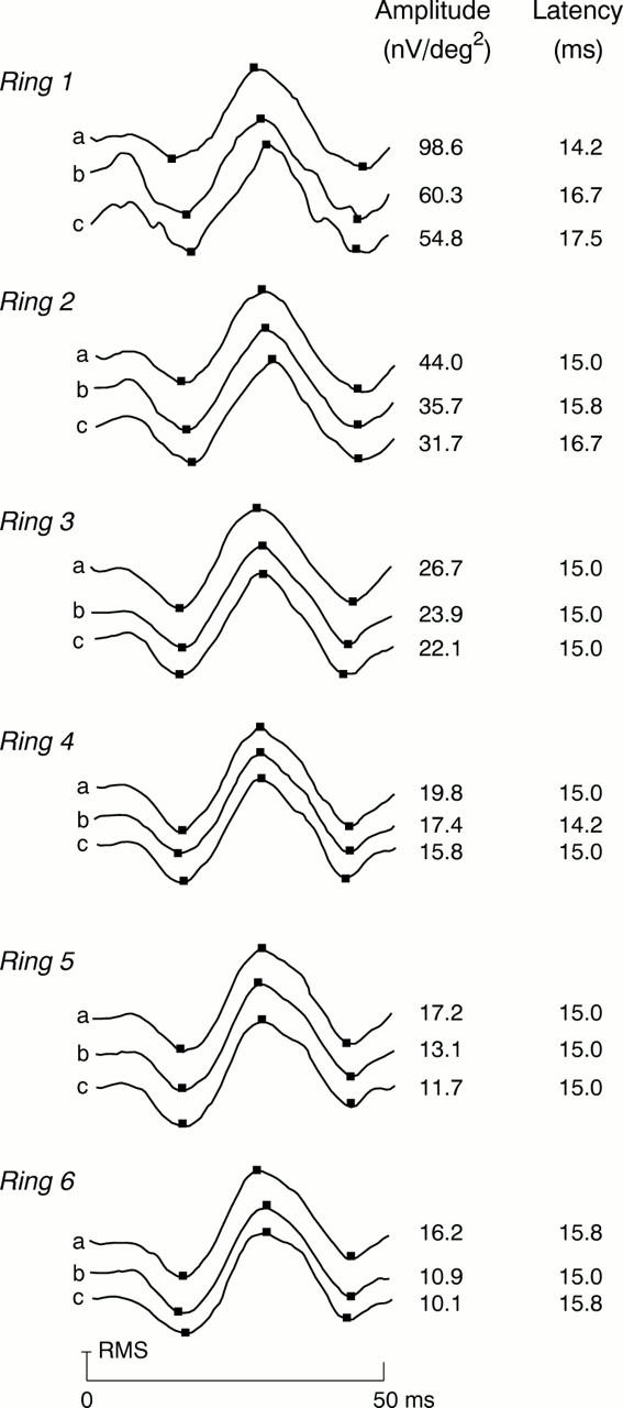
Illustration of the MERG responses and their calculated P1 amplitudes and N1 latencies in (a) normal, (b) fellow, (c) and pre-AMD or early AMD eyes.
Selected References
These references are in PubMed. This may not be the complete list of references from this article.
- Chan H. L., Brown B. Multifocal ERG changes in glaucoma. Ophthalmic Physiol Opt. 1999 Jul;19(4):306–316. doi: 10.1046/j.1475-1313.1999.00439.x. [DOI] [PubMed] [Google Scholar]
- Feeney-Burns L., Burns R. P., Gao C. L. Age-related macular changes in humans over 90 years old. Am J Ophthalmol. 1990 Mar 15;109(3):265–278. doi: 10.1016/s0002-9394(14)74549-0. [DOI] [PubMed] [Google Scholar]
- Fish G. E., Birch D. G. The focal electroretinogram in the clinical assessment of macular disease. Ophthalmology. 1989 Jan;96(1):109–114. doi: 10.1016/s0161-6420(89)32944-7. [DOI] [PubMed] [Google Scholar]
- Fortune B., Schneck M. E., Adams A. J. Multifocal electroretinogram delays reveal local retinal dysfunction in early diabetic retinopathy. Invest Ophthalmol Vis Sci. 1999 Oct;40(11):2638–2651. [PubMed] [Google Scholar]
- Hood D. C., Frishman L. J., Viswanathan S., Robson J. G., Ahmed J. Evidence for a ganglion cell contribution to the primate electroretinogram (ERG): effects of TTX on the multifocal ERG in macaque. Vis Neurosci. 1999 May-Jun;16(3):411–416. doi: 10.1017/s0952523899163028. [DOI] [PubMed] [Google Scholar]
- Hood D. C., Greenstein V., Frishman L., Holopigian K., Viswanathan S., Seiple W., Ahmed J., Robson J. G. Identifying inner retinal contributions to the human multifocal ERG. Vision Res. 1999 Jun;39(13):2285–2291. doi: 10.1016/s0042-6989(98)00296-x. [DOI] [PubMed] [Google Scholar]
- Hosokawa M., Sakagami K., Hongu K., Ohashi Y., Miyamoto F., Ishikawa H. [Use of the multifocal electroretinogram to evaluate retinal function after pars plana vitrectomy for diabetic macular edema]. Nippon Ganka Gakkai Zasshi. 1999 Jun;103(6):464–469. [PubMed] [Google Scholar]
- Kawabata H., Adachi-Usami E. Multifocal electroretinogram in myopia. Invest Ophthalmol Vis Sci. 1997 Dec;38(13):2844–2851. [PubMed] [Google Scholar]
- Kondo M., Miyake Y., Horiguchi M., Suzuki S., Ito Y., Tanikawa A. [Normal values of retinal response densities in multifocal electroretinogram]. Nippon Ganka Gakkai Zasshi. 1996 Oct;100(10):810–816. [PubMed] [Google Scholar]
- Kondo M., Miyake Y., Horiguchi M., Suzuki S., Tanikawa A. Clinical evaluation of multifocal electroretinogram. Invest Ophthalmol Vis Sci. 1995 Sep;36(10):2146–2150. [PubMed] [Google Scholar]
- Kretschmann U., Stilling R., Rüther K., Zrenner E. Familial macular cone dystrophy: diagnostic value of multifocal ERG and two-color threshold perimetry. Graefes Arch Clin Exp Ophthalmol. 1999 May;237(5):429–432. doi: 10.1007/s004170050255. [DOI] [PubMed] [Google Scholar]
- Marmor M. F., Tan F., Sutter E. E., Bearse M. A., Jr Topography of cone electrophysiology in the enhanced S cone syndrome. Invest Ophthalmol Vis Sci. 1999 Jul;40(8):1866–1873. [PubMed] [Google Scholar]
- Mayer M. J., Spiegler S. J., Ward B., Glucs A., Kim C. B. Foveal flicker sensitivity discriminates ARM-risk from healthy eyes. Invest Ophthalmol Vis Sci. 1992 Oct;33(11):3143–3149. [PubMed] [Google Scholar]
- Mayer M. J., Spiegler S. J., Ward B., Glucs A., Kim C. B. Mid-frequency loss of foveal flicker sensitivity in early stages of age-related maculopathy. Invest Ophthalmol Vis Sci. 1992 Oct;33(11):3136–3142. [PubMed] [Google Scholar]
- Piao C. H., Kondo M., Tanikawa A., Terasaki H., Miyake Y. Multifocal electroretinogram in occult macular dystrophy. Invest Ophthalmol Vis Sci. 2000 Feb;41(2):513–517. [PubMed] [Google Scholar]
- Sandberg M. A., Miller S., Gaudio A. R. Foveal cone ERGs in fellow eyes of patients with unilateral neovascular age-related macular degeneration. Invest Ophthalmol Vis Sci. 1993 Nov;34(12):3477–3480. [PubMed] [Google Scholar]
- Seeliger M., Kretschmann U., Apfelstedt-Sylla E., Rüther K., Zrenner E. Multifocal electroretinography in retinitis pigmentosa. Am J Ophthalmol. 1998 Feb;125(2):214–226. doi: 10.1016/s0002-9394(99)80094-4. [DOI] [PubMed] [Google Scholar]
- Si Y. J., Kishi S., Aoyagi K. Assessment of macular function by multifocal electroretinogram before and after macular hole surgery. Br J Ophthalmol. 1999 Apr;83(4):420–424. doi: 10.1136/bjo.83.4.420. [DOI] [PMC free article] [PubMed] [Google Scholar]
- Sutter E. E., Tran D. The field topography of ERG components in man--I. The photopic luminance response. Vision Res. 1992 Mar;32(3):433–446. doi: 10.1016/0042-6989(92)90235-b. [DOI] [PubMed] [Google Scholar]