

Abstract
BACKGROUND/AIM—Scleral search coils are used to measure eye movements. A recent abstract suggests that the coil can affect the eye by decreasing visual acuity, increasing intraocular pressure, and damaging the corneal and conjunctival surface. Such findings, if repeated in all subjects, would cast doubt on the credibility of the search coil as a reliable investigative technique. The aim of this study was to reassess the effect of the scleral search coil on visual function. METHODS—Six volunteer subjects were selected to undergo coil wear and baseline measurements were taken of logMAR visual acuity, non-contact tonometry, keratometry, and slit lamp examination. Four drops of 0.4% benoxinate hydrochloride were instilled before insertion of the lens by an experienced clinician. The lens then remained on the eye for 30 minutes. Measurements of the four ocular health parameters were repeated after 15 and 30 minutes of lens wear. The lens was then removed and the health of the eye reassessed. RESULTS—No obvious pattern of change was found in logMAR visual acuity, keratometry, or intraocular pressure. The lens did produce changes to the conjunctival and corneal surfaces, but this was not considered clinically significant. CONCLUSION—Search coils do not appear to cause any significant effects on visual function. However, thorough prescreening of subjects and post-wear checks should be carried out on all coil wearers to ensure no adverse effects have been caused.
Full Text
The Full Text of this article is available as a PDF (133.4 KB).
Figure 1 .

LogMAR visual acuities (VA) before lens insertion, and at 15 and 30 minutes after lens insertion, for each subject.
Figure 2 .

Mean logMAR visual acuities (VA) before lens insertion, and at 15 and 30 minutes after lens insertion.
Figure 3 .

Individual and mean intraocular pressure (IOP) measurements before and after lens wear.
Figure 4 .

Individual and mean changes in central corneal curvature (K) produced by scleral lens wear.
Figure 5 .
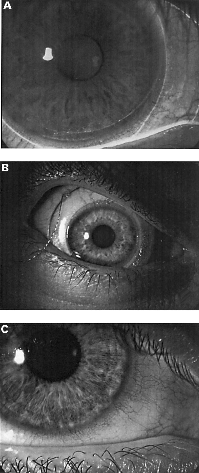
(A) Typical corneal staining produced by scleral lens wear. (B) Position of scleral search coil lens on the eye. (C) Typical limbal redness produced by scleral lens wear.
Selected References
These references are in PubMed. This may not be the complete list of references from this article.
- Barton J. J. Quantitative ocular tests for myasthenia gravis: a comparative review with detection theory analysis. J Neurol Sci. 1998 Feb 18;155(1):104–114. doi: 10.1016/s0022-510x(97)00265-7. [DOI] [PubMed] [Google Scholar]
- Carl J. R., Gellman R. S. Human smooth pursuit: stimulus-dependent responses. J Neurophysiol. 1987 May;57(5):1446–1463. doi: 10.1152/jn.1987.57.5.1446. [DOI] [PubMed] [Google Scholar]
- Collewijn H., Erkelens C. J., Steinman R. M. Binocular co-ordination of human horizontal saccadic eye movements. J Physiol. 1988 Oct;404:157–182. doi: 10.1113/jphysiol.1988.sp017284. [DOI] [PMC free article] [PubMed] [Google Scholar]
- Collewijn H., van der Mark F., Jansen T. C. Precise recording of human eye movements. Vision Res. 1975 Mar;15(3):447–450. doi: 10.1016/0042-6989(75)90098-x. [DOI] [PubMed] [Google Scholar]
- Flipse J. P., Straathof C. S., Van der Steen J., Van Leeuwen A. F., Van Doorn P. A., Van der Meché F. G., Collewijn H. Binocular saccadic eye movements in multiple sclerosis. J Neurol Sci. 1997 May 1;148(1):53–65. doi: 10.1016/s0022-510x(96)05330-0. [DOI] [PubMed] [Google Scholar]
- Millodot M., Owens H. The influence of age on the fragility of the cornea. Acta Ophthalmol (Copenh) 1984 Oct;62(5):819–824. doi: 10.1111/j.1755-3768.1984.tb05810.x. [DOI] [PubMed] [Google Scholar]