Full Text
The Full Text of this article is available as a PDF (295.4 KB).

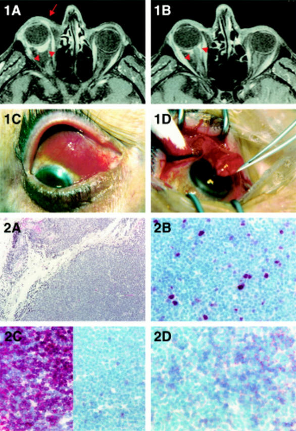
Figures 1 and 2 Clinical and diagnostic presentation of a rare conjunctival MALT lymphoma. (1A) Orbital magnetic resonance imaging (MRI) shows a supraequatorial and epibulbar tumour mass (arrow) with extension to the deep orbit of the right eyeball (arrowheads). (1B) On equatorial sectional plane, MRI revealed tumour masses surrounding the optical nerve (arrowheads). The optical nerve appears not to be infiltrated. (1C and 1D) Excisional biopsy showed a well defined tumour. Histological presentation of conjunctival MALT lymphoma. (2A) Diffuse and homogeneous infiltration of the conjunctival connective tissue by small sized lymphoid cells. Haematoxylin and eosin staining, magnification ×40. (2B) Monoclonal antibody MIB1 recognises a relatively low tumour growth fraction of about 10%. Proliferating cells are indicated by a red nuclear or cytoplasmic signal. APAAP technique, counterstaining by haematoxylin, magnification ×240. (2C) Almost all cells of the tumour mass are immunoreactive for the B cell marker CD79a (left), whereas only few intermingled normal T cells express CD3 (right). Immunoreactive cells are indicated by a red membranous signal. APAAP technique, counterstaining by haematoxylin, magnification ×240. (2D) About 50% of the tumour cells express the T cell antigen CD5. Immunoreactive cells are indicated by a red membranous signal. APAAP technique, counterstaining by haematoxylin, magnification ×240.


