Abstract
AIMS—To calculate the diameter of halos perceived by patients with multifocal intraocular lenses (IOLs) and to stimulate halos in patients with refractive multifocal IOLs in a clinical experiment. METHODS—Calculations were done to show the diameter of halos in the case of the bifocal intraocular lens. 24 patients with a refractive multifocal IOLs and five patients with a monofocal IOL were asked about their subjective observation of halos and were included in a clinical experiment using a computer program (Glare & Halo, FW Fitzke and C Lohmann, Tomey AG) which simulates a light source of 0.15 square degrees (sq deg) in order to stimulate and measure halos. Halo testing took place monoculary, under mesopic conditions through the distance and the near focus of the multifocal lens and through the focus of the monofocal lens. RESULTS—The halo diameter depends on the pupil diameter, the refractive power of the cornea, and distance focus of the multifocal IOL as well as the additional lens power for the near focus. 23 out of 24 patients with a refractive multifocal IOL described halos at night when looking at a bright light source. Only one patient was disturbed by the appearance of halos. Under test conditions, halos were detected in all patients with a refractive multifocal IOL. The halo area testing through the distance focus was 1.05 sq deg ± 0.41, through the near focus 1.07 sq deg ± 0.49 and in the monofocal lens 0.26 sq deg ± 0.13. CONCLUSIONS—Under high contrast conditions halos can be stimulated in all patients with multifocal intraocular lenses. The halo size using the distance or the near focus is identical.
Full Text
The Full Text of this article is available as a PDF (199.1 KB).
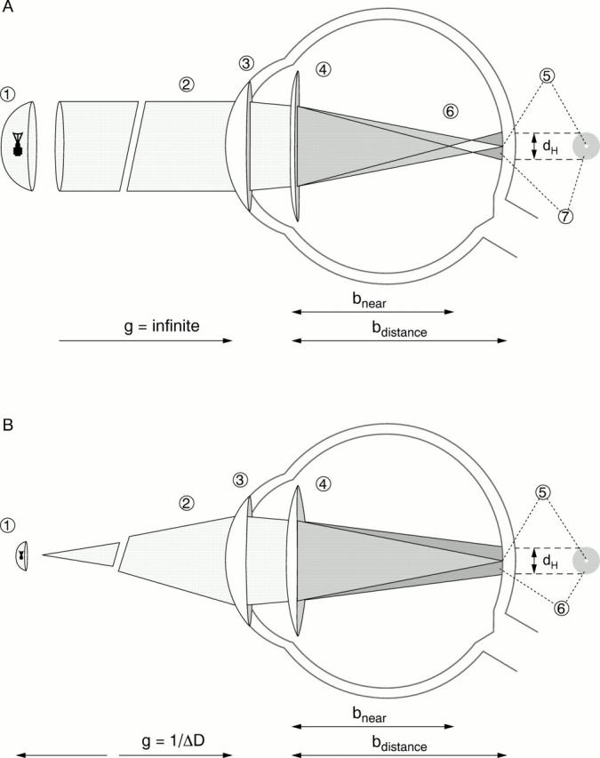
Figure 1 .

(A) A light source nearly at infinity (1) emits parallel light rays (2) that are bent at the cornea (3) and the bifocal lens (4). The distance focus of the bifocal lens produces a sharp image on the retina (5). In the axial presentation this image is shown as a white spot (5). The near focal point in this constellation is in front of the retina (6) producing an out of focus image on the retina (7). The greater diameter of the out of focus image is represented by the dark grey shading in the axial presentation (7) around the focused image (5). (B) A light source is positioned at the reading distance (1) and the light rays reaching the cornea are drawn in (2). These rays are bent at the cornea (3) and the bifocal lens (4). The near focus of the bifocal lens produces a sharp image on the retina (5) also seen in the axial presentation (5). The far focus of the lens would produce an image behind the retina, producing an out of focus image on the retina (6). The greater diameter of this out of focus image is responsible for the halo (6). g = object distance; bnear = image distance produced by the near focus; bdistance = image distance produced by the distance focus; dH = halo diameter; ΔD = power difference between the distance and near focus.
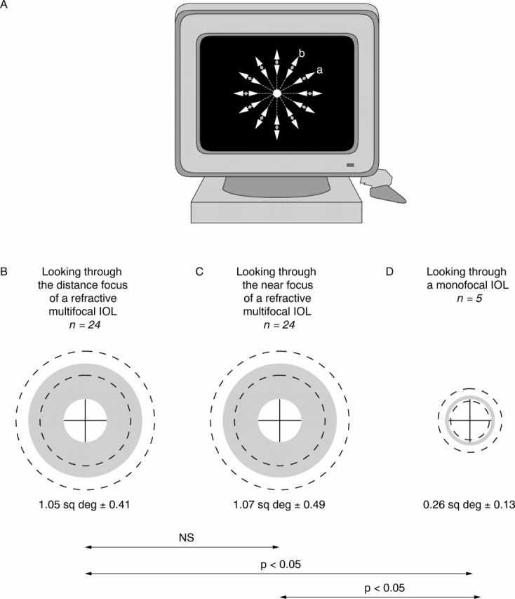
Figure 2 .

(A) The patient is sitting 2 metres away from a video monitor with the best correction. A white, round light source 15 mm in diameter with an illumination of 56.6 cd/m2 is simulated in the centre of the monitor. The background illumination is 0.01 cd/m2. The test takes place under mesopic conditions. A small red spot can be moved with the computer mouse along lines from the periphery to the centre in 30° degree steps (a). The task for the patient is to report when the red spot touches the edge of a possible halo. The halo margin is then determined along the next line 30° away (b). Finally, the extent of the halo is indicated in 30° steps. The computer program automatically computes the area of the halo. (B-D) The circle in the middle represents the extent of the light source. Monocular testing with best distance refraction plus 0.5 D was performed at a distance of 2 metres under mesopic conditions in order to force the patient to use the distance focus (SA 40 N), and with the best distance refraction plus minus 3.0 D in order to force the patient to use the near focus (SA 40 N). In the monofocal group the correction used was the best distance refraction plus 0.5 D (SI 40 NB). The halo is visible as a grey area; the dotted lines represent the standard deviation. ns = not significant.
Selected References
These references are in PubMed. This may not be the complete list of references from this article.
- Arens B., Freudenthaler N., Quentin C. D. Binocular function after bilateral implantation of monofocal and refractive multifocal intraocular lenses. J Cataract Refract Surg. 1999 Mar;25(3):399–404. doi: 10.1016/s0886-3350(99)80089-3. [DOI] [PubMed] [Google Scholar]
- Auffarth G. U., Hunold W., Wesendahl T. A., Mehdorn E. Depth of focus and functional results in patients with multifocal intraocular lenses: a long-term follow-up. J Cataract Refract Surg. 1993 Nov;19(6):685–689. doi: 10.1016/s0886-3350(13)80335-5. [DOI] [PubMed] [Google Scholar]
- Datiles M. B., Gancayco T. Low myopia with low astigmatic correction gives cataract surgery patients good depth of focus. Ophthalmology. 1990 Jul;97(7):922–926. doi: 10.1016/s0161-6420(90)32480-6. [DOI] [PubMed] [Google Scholar]
- Eisenmann D., Hessemer V., Manzke B., Stork W., Jacobi K. W. Modulationsübertragungsfunktion und Kontrastempfindlichkeit refraktiver Mehrzonenmultifokallinsen. Ophthalmologe. 1993 Aug;90(4):343–347. [PubMed] [Google Scholar]
- Eisenmann D., Jacobi F. K., Dick B., Jacobi K. W., Pabst W. Untersuchungen zur Blendempfindlichkeit phaker und pseudophaker Augen. Klin Monbl Augenheilkd. 1996 Feb;208(2):87–92. doi: 10.1055/s-2008-1035175. [DOI] [PubMed] [Google Scholar]
- Eisenmann D., Jacobi K. W. Die ARRAY-Multifokallinse--Funktionsprinzip und klinische Ergebnisse. Klin Monbl Augenheilkd. 1993 Sep;203(3):189–194. doi: 10.1055/s-2008-1045666. [DOI] [PubMed] [Google Scholar]
- Eisenmann D., Jacobi K. W., Krzizok T., Reiner J. Theoretische und klinische Abbildungseigenschaften refraktiver 3-Zonen-Multifokallinsen mit unterschiedlicher Gewichtung von Fern- und Nahfokus. Klin Monbl Augenheilkd. 1994 Nov;205(5):289–297. doi: 10.1055/s-2008-1045531. [DOI] [PubMed] [Google Scholar]
- Eisenmann D., Wagner R., Dick B., Jacobi K. W. Einfluss des Hornhautastigmatismus auf die Kontrastempfindlichkeit bei mono- und multifokaler Pseudophakie--eine theoretische Studie am physikalischen Auge. Klin Monbl Augenheilkd. 1996 Aug-Sep;209(2-3):125–131. doi: 10.1055/s-2008-1035291. [DOI] [PubMed] [Google Scholar]
- Ellingson F. T. Explantation of 3M diffractive intraocular lenses. J Cataract Refract Surg. 1990 Nov;16(6):697–702. doi: 10.1016/s0886-3350(13)81008-5. [DOI] [PubMed] [Google Scholar]
- Goes F. Personal results with the 3M diffractive multifocal intraocular lens. J Cataract Refract Surg. 1991 Sep;17(5):577–582. doi: 10.1016/s0886-3350(13)81044-9. [DOI] [PubMed] [Google Scholar]
- Hessemer V., Frohloff H., Eisenmann D., Jacobi K. W. Mesopisches Sehen bei multi-und, monofokaler Pseudophakie und phaken Kontrollaugen. Ophthalmologe. 1994 Aug;91(4):465–468. [PubMed] [Google Scholar]
- Holladay J. T., Van Dijk H., Lang A., Portney V., Willis T. R., Sun R., Oksman H. C. Optical performance of multifocal intraocular lenses. J Cataract Refract Surg. 1990 Jul;16(4):413–422. doi: 10.1016/s0886-3350(13)80793-6. [DOI] [PubMed] [Google Scholar]
- Jacobi F. K., Kammann J., Jacobi K. W., Grosskopf U., Walden K. Bilateral implantation of asymmetrical diffractive multifocal intraocular lenses. Arch Ophthalmol. 1999 Jan;117(1):17–23. doi: 10.1001/archopht.117.1.17. [DOI] [PubMed] [Google Scholar]
- Jacobi K. W., Eisenmann D. Asymmetrische Mehrzonenlinsen--ein neues Konzept multifokaler Intraokularlinsen. Klin Monbl Augenheilkd. 1993 Apr;202(4):309–314. doi: 10.1055/s-2008-1045597. [DOI] [PubMed] [Google Scholar]
- Lang A., Portney V. Interpreting multifocal intraocular lens modulation transfer functions. J Cataract Refract Surg. 1993 Jul;19(4):505–512. doi: 10.1016/s0886-3350(13)80615-3. [DOI] [PubMed] [Google Scholar]
- Liekfeld A., Pham D. T., Wollensak J. Funktionelle Ergebnisse bei bilateraler Implantation einer faltbaren refraktiven multifokalen Hinterkammerlinse. Klin Monbl Augenheilkd. 1995 Nov;207(5):283–286. doi: 10.1055/s-2008-1035379. [DOI] [PubMed] [Google Scholar]
- Lindstrom R. L. Food and Drug Administration study update. One-year results from 671 patients with the 3M multifocal intraocular lens. Ophthalmology. 1993 Jan;100(1):91–97. [PubMed] [Google Scholar]
- Olsen T., Corydon L. Contrast sensitivity as a function of focus in patients with the diffractive multifocal intraocular lens. J Cataract Refract Surg. 1990 Nov;16(6):703–706. doi: 10.1016/s0886-3350(13)81009-7. [DOI] [PubMed] [Google Scholar]
- Percival S. P., Setty S. S. Prospectively randomized trial comparing the pseudoaccommodation of the AMO ARRAY multifocal lens and a monofocal lens. J Cataract Refract Surg. 1993 Jan;19(1):26–31. doi: 10.1016/s0886-3350(13)80275-1. [DOI] [PubMed] [Google Scholar]
- Pieh S., Weghaupt H., Rainer G., Skorpik C. Sehschärfe und Brillentrageverhalten nach Implantation einer diffraktiven Multifokallinse. Klin Monbl Augenheilkd. 1997 Jan;210(1):38–42. doi: 10.1055/s-2008-1035011. [DOI] [PubMed] [Google Scholar]
- Pieh S., Weghaupt H., Skorpik C. Contrast sensitivity and glare disability with diffractive and refractive multifocal intraocular lenses. J Cataract Refract Surg. 1998 May;24(5):659–662. doi: 10.1016/s0886-3350(98)80261-7. [DOI] [PubMed] [Google Scholar]
- Post C. T., Jr Comparison of depth of focus and low-contrast acuities for monofocal versus multifocal intraocular lens patients at 1 year. Ophthalmology. 1992 Nov;99(11):1658–1664. doi: 10.1016/s0161-6420(92)31735-x. [DOI] [PubMed] [Google Scholar]
- Ravalico G., Baccara F., Rinaldi G. Contrast sensitivity in multifocal intraocular lenses. J Cataract Refract Surg. 1993 Jan;19(1):22–25. doi: 10.1016/s0886-3350(13)80274-x. [DOI] [PubMed] [Google Scholar]
- Reiner J., Speicher L. Eigenschaften durch Injektion erzeugter Intraokularlinsen. Klin Monbl Augenheilkd. 1993 Jan;202(1):49–51. doi: 10.1055/s-2008-1045558. [DOI] [PubMed] [Google Scholar]
- Rossetti L., Carraro F., Rovati M., Orzalesi N. Performance of diffractive multifocal intraocular lenses in extracapsular cataract surgery. J Cataract Refract Surg. 1994 Mar;20(2):124–128. doi: 10.1016/s0886-3350(13)80150-2. [DOI] [PubMed] [Google Scholar]
- Rüther K., Eisenmann D., Zrenner E., Jacobi K. W. Der Einfluss diffraktiver Multifokallinsen auf Kontrastsehen, Gegenlichtsehschärfe und Farbsinn. Klin Monbl Augenheilkd. 1994 Jan;204(1):14–19. doi: 10.1055/s-2008-1035495. [DOI] [PubMed] [Google Scholar]
- Vaquero M., Encinas J. L., Jimenez F. Visual function with monofocal versus multifocal IOLs. J Cataract Refract Surg. 1996 Nov;22(9):1222–1225. doi: 10.1016/s0886-3350(96)80071-x. [DOI] [PubMed] [Google Scholar]
- Walkow T., Liekfeld A., Anders N., Pham D. T., Hartmann C., Wollensak J. A prospective evaluation of a diffractive versus a refractive designed multifocal intraocular lens. Ophthalmology. 1997 Sep;104(9):1380–1386. doi: 10.1016/s0161-6420(97)30127-4. [DOI] [PubMed] [Google Scholar]
- Weghaupt H., Pieh S., Skorpik C. Comparison of pseudoaccommodation and visual quality between a diffractive and refractive multifocal intraocular lens. J Cataract Refract Surg. 1998 May;24(5):663–665. doi: 10.1016/s0886-3350(98)80262-9. [DOI] [PubMed] [Google Scholar]
- Weghaupt H., Pieh S., Skorpik C. Visual properties of the foldable Array multifocal intraocular lens. J Cataract Refract Surg. 1996;22 (Suppl 2):1313–1317. doi: 10.1016/s0886-3350(96)80091-5. [DOI] [PubMed] [Google Scholar]
- Williamson W., Poirier L., Coulon P., Verin P. Compared optical performances of multifocal and monofocal intraocular lenses (contrast sensitivity and dynamic visual acuity) Br J Ophthalmol. 1994 Apr;78(4):249–251. doi: 10.1136/bjo.78.4.249. [DOI] [PMC free article] [PubMed] [Google Scholar]
- Winther-Nielsen A., Corydon L., Olsen T. Contrast sensitivity and glare in patients with a diffractive multifocal intraocular lens. J Cataract Refract Surg. 1993 Mar;19(2):254–257. doi: 10.1016/s0886-3350(13)80952-2. [DOI] [PubMed] [Google Scholar]


