

Full Text
The Full Text of this article is available as a PDF (310.5 KB).
Figure 1 .
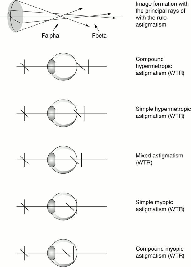
Diagram showing the principal rays in the formation of an astigmatic image. Falpha is the anterior focal point, convergent light rays from the steeper meridian produce a horizontal line focus. Fbeta is the posterior focal point from the flatter meridian producing a vertical line focus. In this example, when Fbeta is coincident with the retina, simple myopic astigmatism with the rule is produced.
Figure 2 .

The ideal cylinder (C) for a given spherical equivalent (SEQ): C = −2SEQ − 0.50 (ie, C = −sphere − 0.25), where SEQ is −0.25 D or less (ie, a myopic correction) and C is a plus cylinder, and C = 2SEQ + 0.50, where SEQ is greater than −0.25 D (ie, a hypermetropic correction).26
Figure 3 .

Residual astigmatism produced when a cylinder axis is shifted away from its correct position: (Csinϕ) DS/−2Csinϕ) DC, axis (θ + 45 + ϕ/2), where C cylinder is set at ϕ angle from the true axis θ. Graphs illustrate how reducing the cylinder power to an "optimal value" can minimise the residual astigmatism. The optimal cylinder power in these circumstances is: Ccos2ϕ, where C is the original full dioptric power of the correcting cylinder, and ϕ is the angle the cylinder is rotated away from the correct position. So if the "optimal value" for the correct cylinder power is used the residual astigmatism is equal to Csin2ϕ, where C is the original full dioptric power of the correcting cylinder and ϕ is the angle the cylinder is rotated away from the correct position.16
Figure 4 .

Diagram demonstrating the principle of vector analysis of the change in the astigmatic refraction following surgery. The arrow direction represents the axis of astigmatism and the length the magnitude. The principle assumes that a theoretical spherocylinder, the surgical induced astigmatism (SIA or CSURG × β°) is "crossed" with the preoperative refraction to produce the postoperative refraction69: SI/CI × θ° + SSURG/CSURG × β° = SR/CR × (θ + ε)°, CSURG = (CI2 + CR2 − 2 CI CR cos2ε), SCYL = (CI + CSURG −C R)/2, sin2β = (CR/CSURG) sin2ε, SSURG = SR − SI − SCYL; CI is the initial or preoperative astigmatism vector at θ° axis (in "plus" cylinder notation), CR is the resultant or postoperative astigmatism vector at θ + ε° axis ("plus" cylinder notation), CSURG is the surgically induced astigmatism vector (SIA, a theoretical construct) at β° axis (in "minus" cylinder notation), SCYL is the spherical equivalent of all the cylindrical components.
Figure 5 .

Diagram demonstrating the principle of decomposition of the astigmatic refraction into x and y values. Cravy56 suggested that cylinder (or astigmatism) of M magnitude at θ° axis maybe characterised by a "y" magnitude at 90° axis (y = M sin θ) and an "x" magnitude at the 0-180° axis (x = M cos θ). Naeser described a single summary value for the astigmatism which he called the polar value of net astigmatism (KP).82 83 85 89-91 This was originally calculated as89: KP = M × (|sin θ| −|cos θ|), but modified later to90: KP = M × (sin2 θ − cos2 θ), where M is the magnitude and θ is the axis of astigmatism, and KP is the polar value referable to the 90° meridian (ie, encompassing the WTR and ATR concept). When sin θ > cos θ, (ie, y > x), the more WTR the astigmatism and the more positive the polar value. Conversely when sin θ < cos θ (ie, y < x) the more ATR the astigmatism and the more negative the polar value.
Figure 6 .

Diagram of the Fourier decomposition of a spherocylindrical lens (S = plano/C = −3.25 × θ = 70). The 360° lens surface power (ie, for all meridians) is represented by the thick line. The three Fourier component terms are: a spherical equivalent (SEQ = S + C/2), a cosine astigmatism term (J × cos2θ), and sine astigmatism term (J × sin2θ) where J = C/2.93
Figure 7 .

Diagram demonstrating the use of the vector representation of the astigmatic refraction to determine the quantities of surgical error. The magnitude error (CERROR) is the simple subtraction of the absolute CSURG magnitude from the absolute CI magnitude in dioptres cylinder. The direction or axis error (ϕ) is the CSURG axis (β) in minus cylinder notation from the CI axis (θ) in plus cylinder notation.34
Figure 8 .
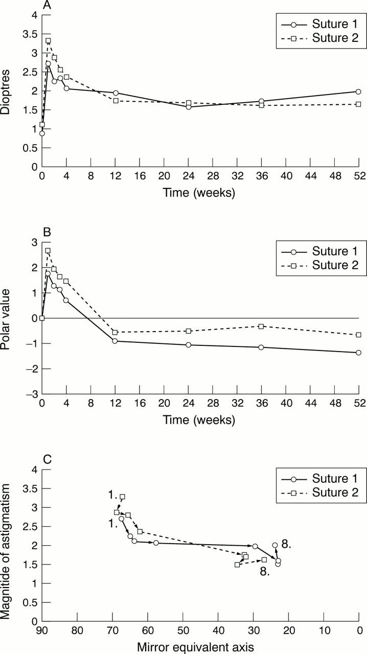
Methods of presenting astigmatism data. The same base data set is used for all the methods of presentation shown in Figures 8, 9, and 10. The data are from two groups of patients who had extracapsular cataract surgery. (A) The change in mean magnitude of astigmatism. (B) The change in the mean Naeser polar value (positive values are with the rule, negative values against the rule). (C) The change in the "expectancy" of astigmatism from 1 week postoperative (no 1) to 12 months postoperatively (no 8) using the "by the rule" or mirror equivalent axis conversion. The "expectancy" is a statistically correct bivariate (or simultaneous) calculation of the magnitude and axis for the grouped astigmatism data (as opposed to the incorrect method of calculating the simple mean of the magnitude or axis independently). This was calculated using the contingency table method of Toulemont.78 The axis values were first converted to mirror equivalent values as shown in Table 2.
Figure 9 .

Methods of presenting astigmatism data. The same base data set is used for all the methods of presentation shown in figures 8, 9, and 10. The data are from two groups of patients who had extracapsular cataract surgery. The change in the "expectancy" of astigmatism regraphed as a balloon plot.
Figure 10 .
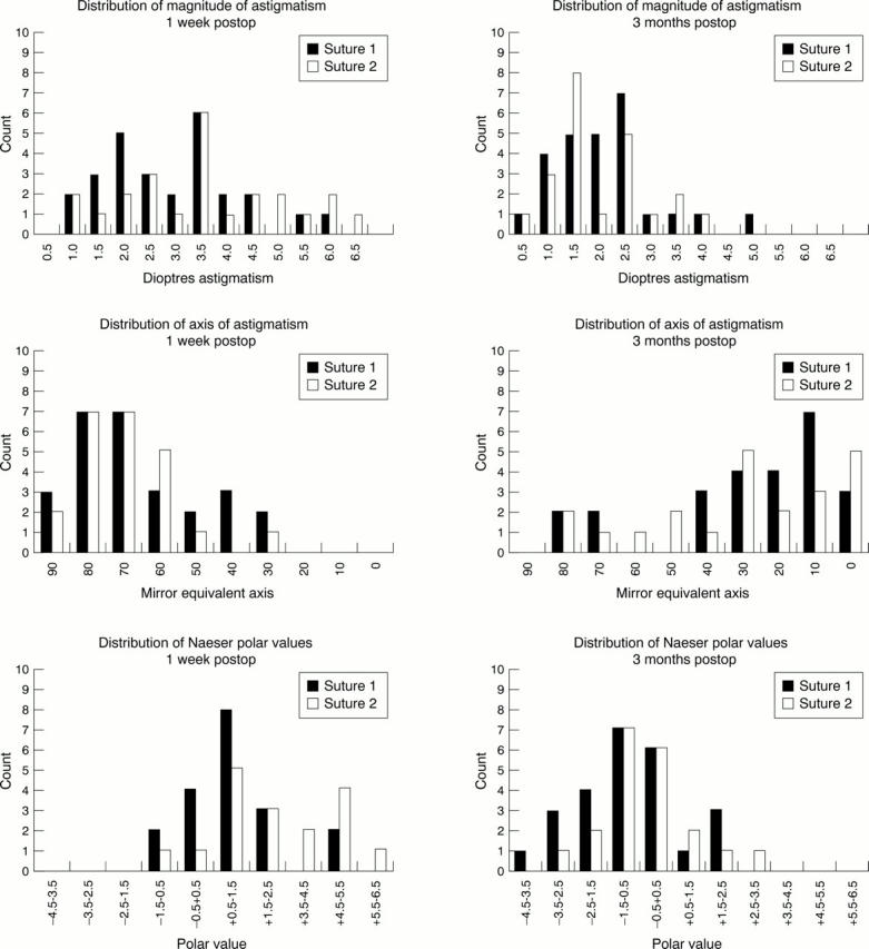
Methods of presenting astigmatism data. The same base data set is used for all the methods of presentation shown in figures 8, 9, and 10. The data are from two groups of patients who had extracapsular cataract surgery. The change in magnitude and axis (converted to mirror equivalent values) of astigmatism and Naeser polar values plotted as frequency histograms at 1 week and 3 months postoperatively.

Selected References
These references are in PubMed. This may not be the complete list of references from this article.
- Alpins N. A. A new method of analyzing vectors for changes in astigmatism. J Cataract Refract Surg. 1993 Jul;19(4):524–533. doi: 10.1016/s0886-3350(13)80617-7. [DOI] [PubMed] [Google Scholar]
- Alpins N. A. New method of targeting vectors to treat astigmatism. J Cataract Refract Surg. 1997 Jan-Feb;23(1):65–75. doi: 10.1016/s0886-3350(97)80153-8. [DOI] [PubMed] [Google Scholar]
- Axt J. C. Longitudinal study of postoperative astigmatism. J Cataract Refract Surg. 1987 Jul;13(4):381–388. doi: 10.1016/s0886-3350(87)80036-6. [DOI] [PubMed] [Google Scholar]
- Binkhorst R. D. The cause of excessive astigmatism with intraocular lens implants. Ophthalmology. 1979 Apr;86(4):672–674. doi: 10.1016/s0161-6420(79)35470-7. [DOI] [PubMed] [Google Scholar]
- Bradbury J. A., Hillman J. S., Cassells-Brown A. Optimal postoperative refraction for good unaided near and distance vision with monofocal intraocular lenses. Br J Ophthalmol. 1992 May;76(5):300–302. doi: 10.1136/bjo.76.5.300. [DOI] [PMC free article] [PubMed] [Google Scholar]
- Buzard K. New formula for vector change in astigmatism. J Cataract Refract Surg. 1993 Nov;19(6):815–816. doi: 10.1016/s0886-3350(13)80366-5. [DOI] [PubMed] [Google Scholar]
- Cravy T. V. Calculation of the change in corneal astigmatism following cataract extraction. Ophthalmic Surg. 1979 Jan;10(1):38–49. [PubMed] [Google Scholar]
- Cravy T. V. Long-term corneal astigmatism related to selected elastic, monofilament, nonabsorbable sutures. J Cataract Refract Surg. 1989 Jan;15(1):61–69. doi: 10.1016/s0886-3350(89)80142-7. [DOI] [PubMed] [Google Scholar]
- Cravy T. V. Routine use of a lateral approach to cataract extraction to achieve rapid and sustained stabilization of postoperative astigmatism. J Cataract Refract Surg. 1991 Jul;17(4):415–423. doi: 10.1016/s0886-3350(13)80848-6. [DOI] [PubMed] [Google Scholar]
- Datiles M. B., Gancayco T. Low myopia with low astigmatic correction gives cataract surgery patients good depth of focus. Ophthalmology. 1990 Jul;97(7):922–926. doi: 10.1016/s0161-6420(90)32480-6. [DOI] [PubMed] [Google Scholar]
- Drews R. C. Astigmatism after cataract surgery: nylon versus Mersilene. Ophthalmic Surg. 1989 Oct;20(10):695–696. [PubMed] [Google Scholar]
- Friedman N. E., Zadnik K., Mutti D. O., Fusaro R. E. Quantifying corneal toricity from videokeratography with Fourier analysis. J Refract Surg. 1996 Jan-Feb;12(1):108–113. doi: 10.3928/1081-597X-19960101-20. [DOI] [PubMed] [Google Scholar]
- GARTNER W. F. ASTIGMATISM AND OPTOMETRIC VECTORS. Am J Optom Arch Am Acad Optom. 1965 Aug;42:459–463. doi: 10.1097/00006324-196508000-00003. [DOI] [PubMed] [Google Scholar]
- Goggin M., Pesudovs K. Assessment of surgically induced astigmatism: toward an international standard II. J Cataract Refract Surg. 1998 Dec;24(12):1552–1553. doi: 10.1016/s0886-3350(98)80339-8. [DOI] [PubMed] [Google Scholar]
- Goggin M., Pesudovs K. Assessment of surgically induced astigmatism: toward an international standard. J Cataract Refract Surg. 1998 Dec;24(12):1548–1550. doi: 10.1016/s0886-3350(98)80337-4. [DOI] [PubMed] [Google Scholar]
- Gross R. H., Miller K. M. Corneal astigmatism after phacoemulsification and lens implantation through unsutured scleral and corneal tunnel incisions. Am J Ophthalmol. 1996 Jan;121(1):57–64. doi: 10.1016/s0002-9394(14)70534-3. [DOI] [PubMed] [Google Scholar]
- Guyton D. L. Prescribing cylinders: the problem of distortion. Surv Ophthalmol. 1977 Nov-Dec;22(3):177–188. doi: 10.1016/0039-6257(77)90054-6. [DOI] [PubMed] [Google Scholar]
- Hall G. W., Campion M., Sorenson C. M., Monthofer S. Reduction of corneal astigmatism at cataract surgery. J Cataract Refract Surg. 1991 Jul;17(4):407–414. doi: 10.1016/s0886-3350(13)80847-4. [DOI] [PubMed] [Google Scholar]
- Harris W. F. Algebra of sphero-cylinders and refractive errors, and their means, variance, and standard deviation. Am J Optom Physiol Opt. 1988 Oct;65(10):794–802. doi: 10.1097/00006324-198810000-00003. [DOI] [PubMed] [Google Scholar]
- Harris W. F. Statistical inference on mean dioptric power: hypothesis testing and confidence regions. Ophthalmic Physiol Opt. 1990 Oct;10(4):363–372. doi: 10.1111/j.1475-1313.1990.tb00883.x. [DOI] [PubMed] [Google Scholar]
- Hjortdal J. O., Erdmann L., Bek T. Fourier analysis of video-keratographic data. A tool for separation of spherical, regular astigmatic and irregular astigmatic corneal power components. Ophthalmic Physiol Opt. 1995 May;15(3):171–185. [PubMed] [Google Scholar]
- Holladay J. T., Cravy T. V., Koch D. D. Calculating the surgically induced refractive change following ocular surgery. J Cataract Refract Surg. 1992 Sep;18(5):429–443. doi: 10.1016/s0886-3350(13)80095-8. [DOI] [PubMed] [Google Scholar]
- Huber C. Myopic astigmatism a substitute for accommodation in pseudophakia. Doc Ophthalmol. 1981 Dec 16;52(2):123–178. doi: 10.1007/BF01675203. [DOI] [PubMed] [Google Scholar]
- Kaye S. B., Campbell S. H., Davey K., Patterson A. A method for assessing the accuracy of surgical technique in the correction of astigmatism. Br J Ophthalmol. 1992 Dec;76(12):738–740. doi: 10.1136/bjo.76.12.738. [DOI] [PMC free article] [PubMed] [Google Scholar]
- Keller P. R., McGhee C. N., Weed K. H. Fourier analysis of corneal topography data after photorefractive keratectomy. J Cataract Refract Surg. 1998 Nov;24(11):1447–1455. doi: 10.1016/s0886-3350(98)80165-x. [DOI] [PubMed] [Google Scholar]
- Koch D. D., Kohnen T., Obstbaum S. A., Rosen E. S. Format for reporting refractive surgical data. J Cataract Refract Surg. 1998 Mar;24(3):285–287. doi: 10.1016/s0886-3350(98)80305-2. [DOI] [PubMed] [Google Scholar]
- Kronish J. W., Forster R. K. Control of corneal astigmatism following cataract extraction by selective suture cutting. Arch Ophthalmol. 1987 Dec;105(12):1650–1655. doi: 10.1001/archopht.1987.01060120048020. [DOI] [PubMed] [Google Scholar]
- Lindstrom R. L. The surgical correction of astigmatism: a clinician's perspective. Refract Corneal Surg. 1990 Nov-Dec;6(6):441–454. [PubMed] [Google Scholar]
- Long D. A., Monica M. L. A prospective evaluation of corneal curvature changes with 3.0- to 3.5-mm corneal tunnel phacoemulsification. Ophthalmology. 1996 Feb;103(2):226–232. doi: 10.1016/s0161-6420(96)30712-4. [DOI] [PubMed] [Google Scholar]
- Masket S. Comparison of suture materials for closure of the scleral pocket incision. J Cataract Refract Surg. 1988 Sep;14(5):548–551. doi: 10.1016/s0886-3350(88)80014-2. [DOI] [PubMed] [Google Scholar]
- Masket S. Keratorefractive aspects of the scleral pocket incision and closure method for cataract surgery. J Cataract Refract Surg. 1989 Jan;15(1):70–77. doi: 10.1016/s0886-3350(89)80143-9. [DOI] [PubMed] [Google Scholar]
- Masket S., Tennen D. G. Astigmatic stabilization of 3.0 mm temporal clear corneal cataract incisions. J Cataract Refract Surg. 1996 Dec;22(10):1451–1455. doi: 10.1016/s0886-3350(96)80146-5. [DOI] [PubMed] [Google Scholar]
- Matthews J. N., Altman D. G., Campbell M. J., Royston P. Analysis of serial measurements in medical research. BMJ. 1990 Jan 27;300(6719):230–235. doi: 10.1136/bmj.300.6719.230. [DOI] [PMC free article] [PubMed] [Google Scholar]
- McDonnell P. J., Moreira H., Clapham T. N., D'Arcy J., Munnerlyn C. R. Photorefractive keratectomy for astigmatism. Initial clinical results. Arch Ophthalmol. 1991 Oct;109(10):1370–1373. doi: 10.1001/archopht.1991.01080100050041. [DOI] [PubMed] [Google Scholar]
- Naeser K. Assessment of surgically induced astigmatism; call for an international standard. J Cataract Refract Surg. 1997 Nov;23(9):1278–1280. doi: 10.1016/s0886-3350(97)80100-9. [DOI] [PubMed] [Google Scholar]
- Naeser K., Behrens J. K. Correlation between polar values and vector analysis. J Cataract Refract Surg. 1997 Jan-Feb;23(1):76–81. doi: 10.1016/s0886-3350(97)80154-x. [DOI] [PubMed] [Google Scholar]
- Naeser K., Behrens J. K., Naeser E. V. Quantitative assessment of corneal astigmatic surgery: expanding the polar values concept. J Cataract Refract Surg. 1994 Mar;20(2):162–168. doi: 10.1016/s0886-3350(13)80158-7. [DOI] [PubMed] [Google Scholar]
- Naeser K. Conversion of keratometer readings to polar values. J Cataract Refract Surg. 1990 Nov;16(6):741–745. doi: 10.1016/s0886-3350(13)81018-8. [DOI] [PubMed] [Google Scholar]
- Naeser K. Format for reporting surgically induced astigmatism on aggregate data. J Cataract Refract Surg. 1998 Dec;24(12):1550–1552. doi: 10.1016/s0886-3350(98)80338-6. [DOI] [PubMed] [Google Scholar]
- Naeser K. Vector analysis may not apply for astigmatic surgery. J Cataract Refract Surg. 1993 Sep;19(5):668–669. doi: 10.1016/s0886-3350(13)80027-2. [DOI] [PubMed] [Google Scholar]
- Naylor E. J. Astigmatic difference in refractive errors. Br J Ophthalmol. 1968 May;52(5):422–425. doi: 10.1136/bjo.52.5.422. [DOI] [PMC free article] [PubMed] [Google Scholar]
- Neumann A. C., McCarty G. R., Sanders D. R., Raanan M. G. Small incisions to control astigmatism during cataract surgery. J Cataract Refract Surg. 1989 Jan;15(1):78–84. doi: 10.1016/s0886-3350(89)80144-0. [DOI] [PubMed] [Google Scholar]
- Novis C. Astigmatism and the toric intraocular lens and other vertex distance effects. Surv Ophthalmol. 1997 Nov-Dec;42(3):268–270. doi: 10.1016/s0039-6257(97)00091-x. [DOI] [PubMed] [Google Scholar]
- Olsen T., Dam-Johansen M., Bek T., Hjortdal J. O. Evaluating surgically induced astigmatism by Fourier analysis of corneal topography data. J Cataract Refract Surg. 1996 Apr;22(3):318–323. doi: 10.1016/s0886-3350(96)80243-4. [DOI] [PubMed] [Google Scholar]
- Olsen T., Dam-Johansen M. Evaluating surgically induced astigmatism. J Cataract Refract Surg. 1994 Sep;20(5):517–522. doi: 10.1016/s0886-3350(13)80231-3. [DOI] [PubMed] [Google Scholar]
- Olsen T. Simple method to calculate the surgically induced refractive change. J Cataract Refract Surg. 1993 Mar;19(2):319–320. doi: 10.1016/s0886-3350(13)80968-6. [DOI] [PubMed] [Google Scholar]
- PETERS H. B. The relationship between refractive error and visual acuity at three age levels. Am J Optom Arch Am Acad Optom. 1961 Apr;38:194–198. doi: 10.1097/00006324-196104000-00002. [DOI] [PubMed] [Google Scholar]
- Parker W. T., Clorfeine G. S. Long-term evolution of astigmatism following planned extracapsular cataract extraction. Arch Ophthalmol. 1989 Mar;107(3):353–357. doi: 10.1001/archopht.1989.01070010363024. [DOI] [PubMed] [Google Scholar]
- Price F. W., Grene R. B., Marks R. G., Gonzales J. S. Astigmatism reduction clinical trial: a multicenter prospective evaluation of the predictability of arcuate keratotomy. Evaluation of surgical nomogram predictability. ARC-T Study Group. Arch Ophthalmol. 1995 Mar;113(3):277–282. doi: 10.1001/archopht.1995.01100030031017. [DOI] [PubMed] [Google Scholar]
- Raasch T. Clinical refraction in three-dimensional dioptric space revisited. Optom Vis Sci. 1997 Jun;74(6):376–380. doi: 10.1097/00006324-199706000-00020. [DOI] [PubMed] [Google Scholar]
- Retzlaff J., Paden P. Y., Ferrell L. Vector analysis of astigmatism. Adding and subtracting spherocylinders. J Cataract Refract Surg. 1993 May;19(3):393–398. doi: 10.1016/s0886-3350(13)80312-4. [DOI] [PubMed] [Google Scholar]
- Richards S. C., Brodstein R. S., Richards W. L., Olson R. J., Combe P. H., Crowell K. E. Long-term course of surgically induced astigmatism. J Cataract Refract Surg. 1988 May;14(3):270–276. doi: 10.1016/s0886-3350(88)80115-9. [DOI] [PubMed] [Google Scholar]
- Rife C. J. More on calculating the surgically induced refractive change: two easy solutions. J Cataract Refract Surg. 1993 Mar;19(2):320–322. doi: 10.1016/s0886-3350(13)80970-4. [DOI] [PubMed] [Google Scholar]
- Rife C. J., Rapuano C. J. Residual error from correcting cylinders. J Cataract Refract Surg. 1993 Nov;19(6):815–815. doi: 10.1016/s0886-3350(13)80365-3. [DOI] [PubMed] [Google Scholar]
- Roper-Hall M. J. Control of astigmatism after surgery and trauma. Br J Ophthalmol. 1982 Sep;66(9):556–559. doi: 10.1136/bjo.66.9.556. [DOI] [PMC free article] [PubMed] [Google Scholar]
- Rowsey J. J., Fouraker B. D. Corneal coupling principles. Int Ophthalmol Clin. 1996 Fall;36(4):29–38. doi: 10.1097/00004397-199603640-00006. [DOI] [PubMed] [Google Scholar]
- SATO T. Posterior incision of cornea; surgical treatment for conical cornea and astigmatism. Am J Ophthalmol. 1950 Jun;33(6):943–948. [PubMed] [Google Scholar]
- Sawusch M. R., Guyton D. L. Optimal astigmatism to enhance depth of focus after cataract surgery. Ophthalmology. 1991 Jul;98(7):1025–1029. doi: 10.1016/s0161-6420(91)32182-1. [DOI] [PubMed] [Google Scholar]
- Schiller G. M., Guthrie D. S. Vector analysis. J Cataract Refract Surg. 1994 May;20(3):366–367. doi: 10.1016/s0886-3350(13)80600-1. [DOI] [PubMed] [Google Scholar]
- Shepherd J. R. Induced astigmatism in small incision cataract surgery. J Cataract Refract Surg. 1989 Jan;15(1):85–88. doi: 10.1016/s0886-3350(89)80145-2. [DOI] [PubMed] [Google Scholar]
- Snibson G. R., Carson C. A., Aldred G. F., Taylor H. R. One-year evaluation of excimer laser photorefractive keratectomy for myopia and myopic astigmatism. Melbourne Excimer Laser Group. Arch Ophthalmol. 1995 Aug;113(8):994–1000. doi: 10.1001/archopht.1995.01100080044028. [DOI] [PubMed] [Google Scholar]
- Steinert R. F., Brint S. F., White S. M., Fine I. H. Astigmatism after small incision cataract surgery. A prospective, randomized, multicenter comparison of 4- and 6.5-mm incisions. Ophthalmology. 1991 Apr;98(4):417–424. [PubMed] [Google Scholar]
- Suzuki R., Tanaka K., Fujiwara N., Kurimoto S. Remarkable postcataract against-the-rule astigmatism after phacoemulsification in 83 of 809 cases. Ann Ophthalmol. 1992 Apr;24(4):147–150. [PubMed] [Google Scholar]
- Thibos L. N., Wheeler W., Horner D. Power vectors: an application of Fourier analysis to the description and statistical analysis of refractive error. Optom Vis Sci. 1997 Jun;74(6):367–375. doi: 10.1097/00006324-199706000-00019. [DOI] [PubMed] [Google Scholar]
- Thornton S. P. Astigmatic keratotomy: a review of basic concepts with case reports. J Cataract Refract Surg. 1990 Jul;16(4):430–435. doi: 10.1016/s0886-3350(13)80795-x. [DOI] [PubMed] [Google Scholar]
- Toulemont P. J. Multivariate analysis versus vector analysis to assess surgically induced astigmatism. J Cataract Refract Surg. 1996 Sep;22(7):977–982. doi: 10.1016/s0886-3350(96)80203-3. [DOI] [PubMed] [Google Scholar]
- Toulemont P. J., Serdarevic O. N. Modèles vectoriels d'analyses des variations d'astigmatisme cornéen antérieur. J Fr Ophtalmol. 1993;16(8-9):472–481. [PubMed] [Google Scholar]
- Trindade F., Oliveira A., Frasson M. Benefit of against-the-rule astigmatism to uncorrected near acuity. J Cataract Refract Surg. 1997 Jan-Feb;23(1):82–85. doi: 10.1016/s0886-3350(97)80155-1. [DOI] [PubMed] [Google Scholar]