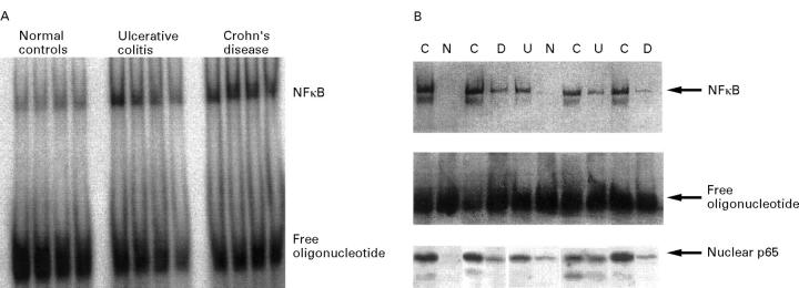
Figure 3 .
Detection of NFκB in the intestinal lamina propria. (A) Nuclear extracts were prepared from colonoscopic biopsy specimens. A representative radioactive electrophoretic mobility shift assay using consensus oligonucleotides to detect NFκB in nuclear extracts from intestinal tissue is shown. In both Crohn's disease (n = 9) and ulcerative colitis (n = 8) increased levels of NFκB were found in comparison with disease specificity controls (n = 5) and healthy volunteers (n = 8). (B) Nuclear extracts were prepared from LPMNCs, which were freshly isolated from colonic biopsy specimens. A blot from a representative non-radioactive gel shift experiment with ten samples (C = Crohn's disease; U = ulcerative colitis; D = disease specificity controls; N = normal controls) is shown. The highest levels of activated NFκB were seen in patients with active Crohn's disease, with similar levels in all four samples. In only one of four normal control samples could low levels of oligonucleotide binding proteins be detected. Levels in patients with ulcerative colitis (four) as well as disease specificity controls (three) appeared to be increased as well, although not as much as in Crohn's disease. The presence of NFκB p65 as part of the complex was controlled by supershift experiments (not shown). The levels of nuclear p65 determined in the same extracts by western blot are shown in the lower part of the figure.

