

Abstract
In a newborn girl with a history of connatal liver damage, histological examination of a liver biopsy sample taken during the seventh week of life revealed incipient destruction of bile ducts. Very high titres of antimitochondrial antibodies were later detected in the plasma. As the hepatic injury tended towards fibrosis, the histological diagnosis became primary biliary cirrhosis. Autoantibodies against E1α, E2, and E3 subunits and protein X component of pyruvate dehydrogenase complex, and against citrate synthase were detected on western immunoblotting in a 1 in 1000 dilution of the patient's serum. The patient died of her illness at 11 years of age. In liver specimens obtained at autopsy human immunoglobulin deposition was detected on the surface of almost all hepatic cells by immunohistology. As there is a physical and functional interaction between pyruvate dehydrogenase and citrate synthase within the mitochondria, the presence of autoantibodies against certain proteins in the patient suggests that in this form of the disease the molecular recognition and then the autoimmunisation process could be directed against a mitochondrial enzyme cluster containing both pyruvate dehydrogenase and citrate synthase.
Keywords: primary biliary cirrhosis; pyruvate dehydrogenase; citrate synthase
Full Text
The Full Text of this article is available as a PDF (192.7 KB).
Figure 1 .
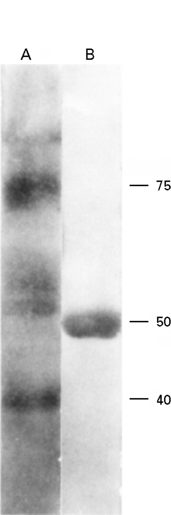
Autoantibodies against PDC and CS. Lane A, from the top: E2 (apparent molecular weight 70-74 kDa), protein X (50-56 kDa), E3 (55 kDa), and E1α (41 kDa) subunits of PDC. Lane B: western blotting for CS (50 kDa) showed anticitrate synthase antibody in the serum.
Figure 2 .
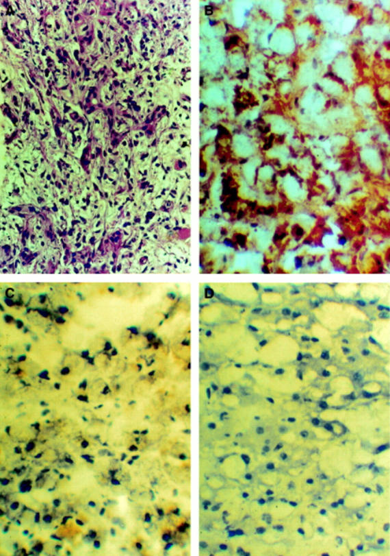
(A) In needle biopsy samples signs of biliary cirrhosis were prominent at six years of age (haematoxylin-eosin staining). (B) Following immunostaining of the liver there was intensive reaction with antihuman immunoglobulin predominantly on the surface of the hepatocytes. (C) Hepatocytes did not show binding of antimouse immunoglobulins (haematoxylin nuclear staining background; the yellow areas are from the bile pigments). (D) The hepatocytes of an adult patient who had died of alcoholic cirrhosis did not bind human immunoglobulins.