Abstract
Background—Flat adenomas are non-exophytic with a flat top or central depression and histologically the depth of dysplastic tissue is never more than twice the mucosal thickness. Flat adenomas frequently contain severely dysplastic tissue, and may progress rapidly through the adenoma-carcinoma sequence. Flat lesions have never been described in a British asymptomatic population. Aims—To determine whether flat adenomas exist in an asymptomatic population participating in a large randomised controlled trial of flexible sigmoidoscopy screening. Patients—A total of 3000 subjects (aged 55-64 years) underwent screening by flexible sigmoidoscopy. Methods—All polyps were removed and sent for histology. The number of polyps with endoscopic and histological features of flat adenomas was recorded. Results—Three subjects had a total of four flat lesions—that is, one per 1000 people screened. Three contained severely dysplastic tissue, one a focus of adenocarcinoma. Three of the four lesions were less than 5 mm in size and the fourth was 15 mm in diameter. Conclusions—Flat lesions with severe dysplasia exist in the asymptomatic population. This has major implications for gastroenterologists who should be trained to identify them. Their existence is of importance to molecular biologists and epidemiologists investigating the aetiology of colorectal cancer.
Keywords: flat adenomas; screening; flexible sigmoidoscopy
Full Text
The Full Text of this article is available as a PDF (89.2 KB).
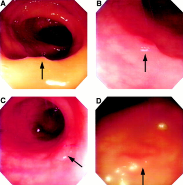
Figure 1 .
Endoscopic appearance of flat lesions from patient A (A), patient B (B,C), and patient C (D).
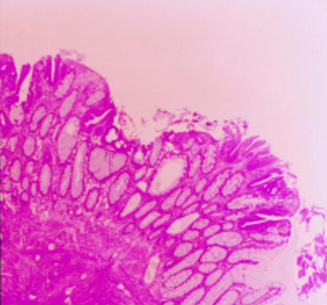
Figure 2 .
Low power view of histology from patient C.
Selected References
These references are in PubMed. This may not be the complete list of references from this article.
- Atkin W. S., Cuzick J., Northover J. M., Whynes D. K. Prevention of colorectal cancer by once-only sigmoidoscopy. Lancet. 1993 Mar 20;341(8847):736–740. doi: 10.1016/0140-6736(93)90499-7. [DOI] [PubMed] [Google Scholar]
- Kudo S., Tamure S., Nakajima T., Hirota S., Asano M., Ito O., Kusaka H. Depressed type of colorectal cancer. Endoscopy. 1995 Jan;27(1):54–61. doi: 10.1055/s-2007-1005633. [DOI] [PubMed] [Google Scholar]
- Mitooka H., Fujimori T., Maeda S., Nagasako K. Minute flat depressed neoplastic lesions of the colon detected by contrast chromoscopy using an indigo carmine capsule. Gastrointest Endosc. 1995 May;41(5):453–459. doi: 10.1016/s0016-5107(05)80003-3. [DOI] [PubMed] [Google Scholar]
- Muto T., Bussey H. J., Morson B. C. The evolution of cancer of the colon and rectum. Cancer. 1975 Dec;36(6):2251–2270. doi: 10.1002/cncr.2820360944. [DOI] [PubMed] [Google Scholar]
- Muto T., Kamiya J., Sawada T., Konishi F., Sugihara K., Kubota Y., Adachi M., Agawa S., Saito Y., Morioka Y. Small "flat adenoma" of the large bowel with special reference to its clinicopathologic features. Dis Colon Rectum. 1985 Nov;28(11):847–851. doi: 10.1007/BF02555490. [DOI] [PubMed] [Google Scholar]
- Stolte M., Bethke B. Colorectal mini-de novo carcinoma: a reality in Germany too. Endoscopy. 1995 May;27(4):286–290. doi: 10.1055/s-2007-1005694. [DOI] [PubMed] [Google Scholar]
- Stryker S. J., Wolff B. G., Culp C. E., Libbe S. D., Ilstrup D. M., MacCarty R. L. Natural history of untreated colonic polyps. Gastroenterology. 1987 Nov;93(5):1009–1013. doi: 10.1016/0016-5085(87)90563-4. [DOI] [PubMed] [Google Scholar]
- Williams A. C., Harper S. J., Paraskeva C. Neoplastic transformation of a human colonic epithelial cell line: in vitro evidence for the adenoma to carcinoma sequence. Cancer Res. 1990 Aug 1;50(15):4724–4730. [PubMed] [Google Scholar]
- Wolber R. A., Owen D. A. Flat adenomas of the colon. Hum Pathol. 1991 Jan;22(1):70–74. doi: 10.1016/0046-8177(91)90064-v. [DOI] [PubMed] [Google Scholar]


