Abstract
BACKGROUND—Helical computed tomography performed after intravenous administration of a cholangiographic contrast material (HCT-cholangiography) may be useful for detecting bile duct stones in non-jaundiced patients. However, this method has never been compared with other non-invasive biliary imaging tests. AIMS—To compare prospectively HCT-cholangiography and endosonography (EUS) in a group of non-jaundiced patients with suspected bile duct stones. METHODS—Fifty two subjects underwent both HCT-cholangiography and EUS. Endoscopic retrograde cholangiography (ERCP), with or without instrumental bile duct exploration, served as a reference method, and was successful in all but two patients. RESULTS—Thirty four patients (68%) were found to have choledocholithiasis at ERCP. The sensitivity for HCT-cholangiography in stone detection was 85%, specificity 88%, and accuracy 86%. For EUS the sensitivity was 91%, specificity 100%, and accuracy 94%. The differences were not significant. No serious complications occurred with either method. CONCLUSIONS—HCT-cholangiography and EUS are safe and comparably accurate methods for detecting bile duct stones in non-jaundiced patients. Keywords: bile duct; calculi; endoscopic ultrasonography; computed tomography; cholangiography.
Full Text
The Full Text of this article is available as a PDF (139.8 KB).
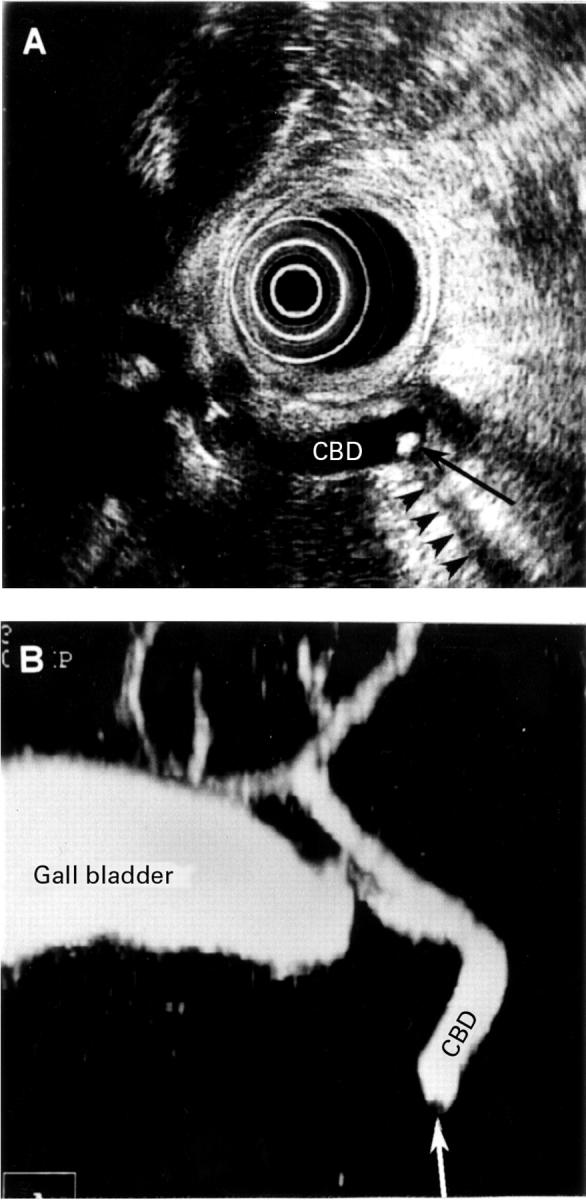
Figure 1 .

Small stone, 4 mm in diameter, in the distal part of undilated common bile duct (CBD). (A) Endosonographic image showing the stone (black arrow) with acoustic shadowing (arrowheads). (B) Helical computed tomographic cholangiography; maximum intensity projection image shows the gall bladder and both the intrahepatic and extrahepatic bile ducts. The small filling defect (white arrow) close to the end of the common bile duct represents the stone.
Figure 2 .

A single 10 mm bile duct stone shown by endosonography (A) and helical computed tomographic (HCT)-cholangiography (B, C). (A) A hyperechoic structure (white arrow) is seen associated with acoustic shadowing (black arrowheads), which is a typical appearance of a stone at endosonography; CBD indicates the common bile duct. (B) HCT-cholangiography; an axial image at the level of the distal common bile duct shows a filling defect (black arrow) which corresponds to the stone seen at endosonography. White arrowheads delineate the cross section of the common bile duct containing contrast enhanced bile. (C) HCT-cholangiography; multiplanar reconstruction image in the same patient. The stone (white arrow) is seen as a polygonal filling defect.
Selected References
These references are in PubMed. This may not be the complete list of references from this article.
- Amouyal P., Amouyal G., Lévy P., Tuzet S., Palazzo L., Vilgrain V., Gayet B., Belghiti J., Fékété F., Bernades P. Diagnosis of choledocholithiasis by endoscopic ultrasonography. Gastroenterology. 1994 Apr;106(4):1062–1067. doi: 10.1016/0016-5085(94)90768-4. [DOI] [PubMed] [Google Scholar]
- Aubertin J. M., Levoir D., Bouillot J. L., Becheur H., Bloch F., Aouad K., Alexandre J. H., Petite J. P. Endoscopic ultrasonography immediately prior to laparoscopic cholecystectomy: a prospective evaluation. Endoscopy. 1996 Oct;28(8):667–673. doi: 10.1055/s-2007-1005574. [DOI] [PubMed] [Google Scholar]
- Barkun A. N., Barkun J. S., Fried G. M., Ghitulescu G., Steinmetz O., Pham C., Meakins J. L., Goresky C. A. Useful predictors of bile duct stones in patients undergoing laparoscopic cholecystectomy. McGill Gallstone Treatment Group. Ann Surg. 1994 Jul;220(1):32–39. doi: 10.1097/00000658-199407000-00006. [DOI] [PMC free article] [PubMed] [Google Scholar]
- Baron R. L. Common bile duct stones: reassessment of criteria for CT diagnosis. Radiology. 1987 Feb;162(2):419–424. doi: 10.1148/radiology.162.2.3797655. [DOI] [PubMed] [Google Scholar]
- Baron R. L. Diagnosing choledocholithiasis: how far can we push helical CT? Radiology. 1997 Jun;203(3):601–603. doi: 10.1148/radiology.203.3.9169674. [DOI] [PubMed] [Google Scholar]
- Bearcroft P. W., Lomas D. J. Magnetic resonance cholangiopancreatography. Gut. 1997 Aug;41(2):135–137. doi: 10.1136/gut.41.2.135. [DOI] [PMC free article] [PubMed] [Google Scholar]
- Canto M. I., Chak A., Stellato T., Sivak M. V., Jr Endoscopic ultrasonography versus cholangiography for the diagnosis of choledocholithiasis. Gastrointest Endosc. 1998 Jun;47(6):439–448. doi: 10.1016/s0016-5107(98)70242-1. [DOI] [PubMed] [Google Scholar]
- Chan A. C., Chung S. C., Wyman A., Kwong K. H., Ng E. K., Lau J. Y., Lau W. Y., Lai C. W., Sung J. J., Li A. K. Selective use of preoperative endoscopic retrograde cholangiopancreatography in laparoscopic cholecystectomy. Gastrointest Endosc. 1996 Mar;43(3):212–215. doi: 10.1016/s0016-5107(96)70318-8. [DOI] [PubMed] [Google Scholar]
- Chan Y. L., Chan A. C., Lam W. W., Lee D. W., Chung S. S., Sung J. J., Cheung H. S., Li A. K., Metreweli C. Choledocholithiasis: comparison of MR cholangiography and endoscopic retrograde cholangiography. Radiology. 1996 Jul;200(1):85–89. doi: 10.1148/radiology.200.1.8657949. [DOI] [PubMed] [Google Scholar]
- Cotton P. B. Endoscopic retrograde cholangiopancreatography and laparoscopic cholecystectomy. Am J Surg. 1993 Apr;165(4):474–478. doi: 10.1016/s0002-9610(05)80944-4. [DOI] [PubMed] [Google Scholar]
- Goldberg H. I. Helical cholangiography: complementary or substitute study for endoscopic retrograde cholangiography? Radiology. 1994 Sep;192(3):615–616. doi: 10.1148/radiology.192.3.8058922. [DOI] [PubMed] [Google Scholar]
- Holzknecht N., Gauger J., Sackmann M., Thoeni R. F., Schurig J., Holl J., Weinzierl M., Helmberger T., Paumgartner G., Reiser M. Breath-hold MR cholangiography with snapshot techniques: prospective comparison with endoscopic retrograde cholangiography. Radiology. 1998 Mar;206(3):657–664. doi: 10.1148/radiology.206.3.9494483. [DOI] [PubMed] [Google Scholar]
- Klein H. M., Wein B., Truong S., Pfingsten F. P., Günther R. W. Computed tomographic cholangiography using spiral scanning and 3D image processing. Br J Radiol. 1993 Sep;66(789):762–767. doi: 10.1259/0007-1285-66-789-762. [DOI] [PubMed] [Google Scholar]
- Kwon A. H., Inui H., Imamura A., Uetsuji S., Kamiyama Y. Preoperative assessment for laparoscopic cholecystectomy: feasibility of using spiral computed tomography. Ann Surg. 1998 Mar;227(3):351–356. doi: 10.1097/00000658-199803000-00006. [DOI] [PMC free article] [PubMed] [Google Scholar]
- Kwon A. H., Uetsuji S., Ogura T., Kamiyama Y. Spiral computed tomography scanning after intravenous infusion cholangiography for biliary duct anomalies. Am J Surg. 1997 Oct;174(4):396–402. [PubMed] [Google Scholar]
- Lindsey I., Nottle P. D., Sacharias N. Preoperative screening for common bile duct stones with infusion cholangiography: review of 1000 patients. Ann Surg. 1997 Aug;226(2):174–178. doi: 10.1097/00000658-199708000-00009. [DOI] [PMC free article] [PubMed] [Google Scholar]
- Neitlich J. D., Topazian M., Smith R. C., Gupta A., Burrell M. I., Rosenfield A. T. Detection of choledocholithiasis: comparison of unenhanced helical CT and endoscopic retrograde cholangiopancreatography. Radiology. 1997 Jun;203(3):753–757. doi: 10.1148/radiology.203.3.9169700. [DOI] [PubMed] [Google Scholar]
- Nilsson U. Adverse reactions to iotroxate at intravenous cholangiography. A prospective clinical investigation and review of the literature. Acta Radiol. 1987 Sep-Oct;28(5):571–575. [PubMed] [Google Scholar]
- Palazzo L., Girollet P. P., Salmeron M., Silvain C., Roseau G., Canard J. M., Chaussade S., Couturier D., Paolaggi J. A. Value of endoscopic ultrasonography in the diagnosis of common bile duct stones: comparison with surgical exploration and ERCP. Gastrointest Endosc. 1995 Sep;42(3):225–231. doi: 10.1016/s0016-5107(95)70096-x. [DOI] [PubMed] [Google Scholar]
- Prat F., Amouyal G., Amouyal P., Pelletier G., Fritsch J., Choury A. D., Buffet C., Etienne J. P. Prospective controlled study of endoscopic ultrasonography and endoscopic retrograde cholangiography in patients with suspected common-bileduct lithiasis. Lancet. 1996 Jan 13;347(8994):75–79. doi: 10.1016/s0140-6736(96)90208-1. [DOI] [PubMed] [Google Scholar]
- Reinhold C., Taourel P., Bret P. M., Cortas G. A., Mehta S. N., Barkun A. N., Wang L., Tafazoli F. Choledocholithiasis: evaluation of MR cholangiography for diagnosis. Radiology. 1998 Nov;209(2):435–442. doi: 10.1148/radiology.209.2.9807570. [DOI] [PubMed] [Google Scholar]
- Shim C. S., Joo J. H., Park C. W., Kim Y. S., Lee J. S., Lee M. S., Hwang S. G. Effectiveness of endoscopic ultrasonography in the diagnosis of choledocholithiasis prior to laparoscopic cholecystectomy. Endoscopy. 1995 Aug;27(6):428–432. doi: 10.1055/s-2007-1005735. [DOI] [PubMed] [Google Scholar]
- Stockberger S. M., Wass J. L., Sherman S., Lehman G. A., Kopecky K. K. Intravenous cholangiography with helical CT: comparison with endoscopic retrograde cholangiography. Radiology. 1994 Sep;192(3):675–680. doi: 10.1148/radiology.192.3.8058932. [DOI] [PubMed] [Google Scholar]
- Sugiyama M., Atomi Y. Endoscopic ultrasonography for diagnosing choledocholithiasis: a prospective comparative study with ultrasonography and computed tomography. Gastrointest Endosc. 1997 Feb;45(2):143–146. doi: 10.1016/s0016-5107(97)70237-2. [DOI] [PubMed] [Google Scholar]
- Zidi S. H., Prat F., Le Guen O., Rondeau Y., Rocher L., Fritsch J., Choury A. D., Pelletier G. Use of magnetic resonance cholangiography in the diagnosis of choledocholithiasis: prospective comparison with a reference imaging method. Gut. 1999 Jan;44(1):118–122. doi: 10.1136/gut.44.1.118. [DOI] [PMC free article] [PubMed] [Google Scholar]


