Abstract
OBJECTIVE—Laser Doppler flowmetry (LDF) has been used as a research tool to measure splanchnic perfusion. In this report, we aim to demonstrate the clinical value of this technique in perioperative monitoring of transplanted small bowel. METHODS—A 24 year old man underwent small bowel transplantation using a previously described technique. Microvascular blood flow in the transplanted bowel was measured using an LDF splanchnic probe. Postoperatively this was applied to the stoma facilitating direct measurements of graft mucosal flow. Measurements (perfusion units (PU)) were documented prior to implantation, post-reperfusion, postoperatively, during graft ischaemia, and in response to therapeutic interventions (dopexamine and phenylephrine infusions). RESULTS—Prior to transplantation, biological zero was established. Flow at five, 15, and 60 minutes after reperfusion was 74 (1.9) PU, 87.5 (3.3) PU, and 141.5 (2.5) PU, respectively. Postoperative mucosal flow was 141.6 (2.9) PU. Subsequent LDF measurement detected absence of flow even though clinical signs suggested only moderate reduction. This was confirmed on surgical re-exploration and facilitated prompt resection of a non-viable segment. Fluid and dopexamine administration resulted in a dose dependent improvement in flow, independent of blood pressure. Addition of phenylephrine increased total mucosal flow and unmasked a cyclical component. CONCLUSION—This case demonstrates the clinical value of LDF as an "alarm" to indicate graft perfusion failure and as a monitor for therapeutic interventions. Phenylephrine and dopexamine may both be of value in improving mucosal flow in the transplanted small bowel. Keywords: laser Doppler; small bowel transplantation; mucosa; dopexamine; flow motion phenomenon
Full Text
The Full Text of this article is available as a PDF (132.2 KB).
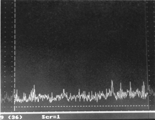
Figure 1 .
Laser Doppler flowmetry (LDF) record (in perfusion units/time) of graft microvascular blood flow at baseline.
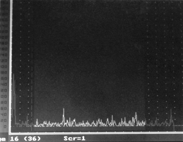
Figure 2 .
Laser Doppler flowmetry (LDF) record at the time of critical graft ischaemia. Blood flow approaches previously determined biological zero (horizontal dashed line indicates biological zero flow).
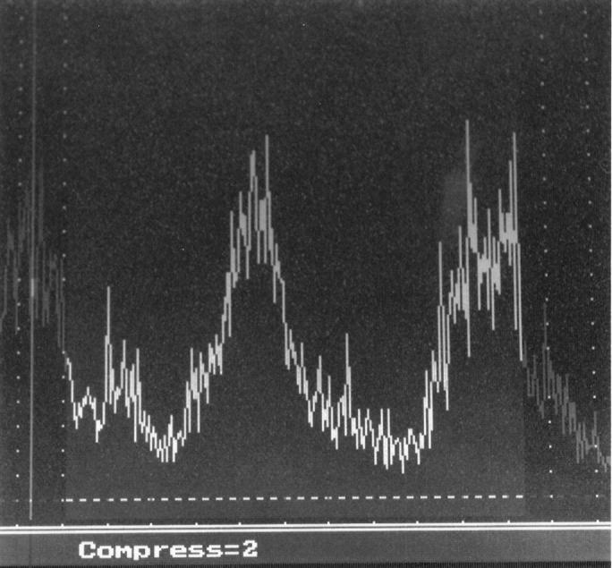
Figure 3 .

Laser Doppler flowmetry (LDF) record following administration of phenylephrine. In addition to an overall increase in graft mucosal blood flow, there is the appearance of a marked cyclical component.


