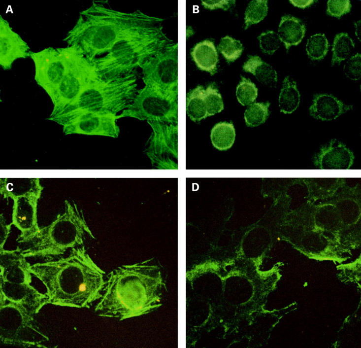
Figure 1 .

Actin stress fibre immunostaining pattern in indirect immunofluorescence of HEp-2 cells using serum from patient No 21 (see table 1), at a screening dilution of 1:40, and the FITC conjugate antihuman IgA (photomicrograph images, ×50; (A)). Typical actin stress fibres appear as long cytoplasmic parallel filaments. (B) Negative immunostaining image obtained using monoclonal antitransglutaminase antibody on HEp-2 cells (photomicrograph images, ×50). The results of actin absorption experiments using 1:1000 diluted serum are shown in (C) (before absorption) and (D) (after absorption).
