Abstract
We report the clinicopathological findings of two patients with ectopic gastric mucosa within the gall ladder. The first patient, a 78 year old man, was asymptomatic. He was admitted to hospital for a colon adenocarcinoma. Intraoperatively, a firm nodule was palpable in the gall bladder. Histological examination of the resected specimen revealed a body type gastric mucosa in the submucosa, adjacent to which were extensive pyloric gland and intestinal metaplasia with mild to moderate dysplasia. The remaining gall bladder mucosa demonstrated changes of chronic cholecystitis. The second patient was a 62 year old woman with symptoms of chronic cholecystitis. The preoperative diagnosis was consistent with this diagnosis with a "polyp" at the junction of the neck and cystic duct. Cholecystectomy was performed and the histological examination of the resected specimen showed that the "polyp" consisted of heterotopic gastric mucosa with glands of body and fundus type. In the remaining mucosa, chronic cholecystitis was evident. To the best of our knowledge, this is the first report of a clinicopathological presentation of heterotopic gastric mucosa, pyloric gland type, and intestinal metaplasia with dysplastic changes in the gall bladder. As heterotopic tissue may promote carcinogenesis of the gall bladder, close attention should be paid to any occurrence of such lesions in this anatomical region. Keywords: heterotopic gastric mucosa; gall bladder; intestinal metaplasia; dysplasia; precancerous lesion
Full Text
The Full Text of this article is available as a PDF (175.1 KB).
Figure 1 .
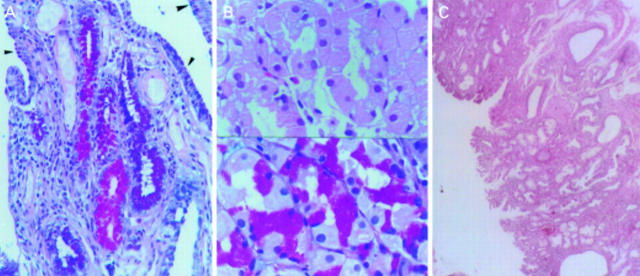
(A) Alcian blue-periodic acid-Schiff (PAS) stain in normal (arrowheads) and metaplastic gall bladder epithelium. The acid mucins of intestinal metaplasia are blue whereas neutral mucins of gastric metaplasia (pyloric type) are red (×320). (B) Heterotopic gastric mucosa of body type in the gall bladder; parietal cells (top) have a large pyramidal shape and plump eosinophilic cytoplasm (haematoxylin-eosin ×480); same area (bottom) stained with Alcian blue-PAS (×480). (C) Polyp of the gall bladder consisting of heterotopic gastric mucosa (haematoxylin-eosin ×180).
Figure 2 .
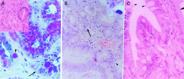
(A) Mild and focally moderately dysplastic epithelium (arrow), occurring in intestinal metaplasia (arrowheads) (Alcian blue-periodic acid-Schiff (PAS) stain ×320) (inset: mild dysplasia developing in pyloric type metaplastic epithelium (haematoxylin-eosin ×320). (B) In this field the transition between normal (arrowhead) and metaplastic epithelium (arrow) can be seen (haematoxylin-eosin ×320); mild dysplasia is focally well discerned (inset: haematoxylin-eosin ×850). (C) Intestinal metaplasia of a tubule (arrowheads) in close proximity to the normal gall bladder epithelium (haematoxylin-eosin ×540).


