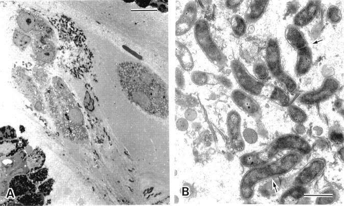
Figure 9 .
Electron microscopic appearance of the aggregates of Helicobacter pylori within the surface mucous gel layer (SMGL). (A) The aggregates of H pylori located in the reticular pattern layer were accompanied by degenerated epithelial cells (bar=10 µm). (B) At higher magnification H pylori showed the typical spiral form with well developed flagellae. Division of the organisms was seen (arrows). Around the microcolonies, the electron density of the SMGL was considerably decreased and the reticular pattern became obscured (see fig 4) (bar=1 µm).

