Abstract
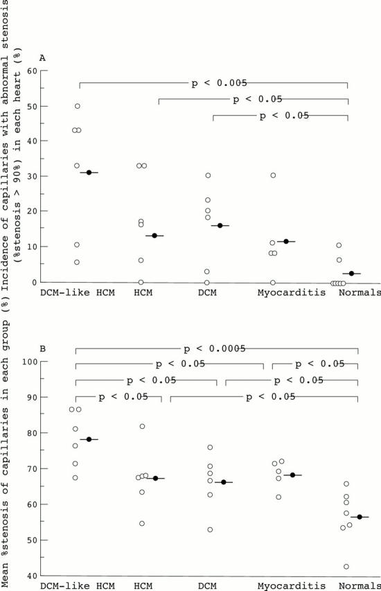
Background—Abnormal microcirculation has been suggested in hearts with pathological conditions, particularly in hypertrophic hearts, even in the presence of normal epicardial coronary arteries. However, the morphology of coronary capillaries has not been well investigated in those hearts. Methods—Ultrastructural morphometry of the capillaries in 47 endomyocardial biopsy specimens taken from 30 patients was performed. Patients—Six patients had hypertrophic cardiomyopathy with dilated cardiomyopathy-like features (DCM-like HCM), six had HCM, six had DCM, five had postmyocarditis, and seven were normal subjects. Results—The short axial diameters of capillaries were similar among the groups. Abnormal stenosis of more than 90% luminal narrowing was found in 31% of capillaries of the DCM-like HCM group, 16% of the HCM group, 13% of the DCM group, 11% of the postmyocarditis group, and 2% of the normal subjects. Mean (SD) stenosis of the lumen was most severe in DCM-like HCM (78(8)%), and more severe in HCM (67(9)%), DCM (66(8)%), and postmyocarditis (68(4)%) than normal subjects (56(8)%). The mean cross sectional areas of capillaries were similar among the groups; however, the endothelial cellular area was significantly (p < 0.05) greater in DCM-like HCM (24.2 (8.2) µm2) than in normal subjects (14.7 (1.8) µm2), indicating that capillary narrowing was due to the increased volume of capillary endothelial cells. The endothelial cells of the stenosed capillaries showed severely oedematous changes of the cytoplasm wholely or partially, but the cytoplasmic organelles and nuclei appeared intact. Conclusion—Narrowing of the coronary capillaries may be of pathophysiological significance in microcirculatory abnormality in hypertrophic hearts, particularly in patients with DCM-like HCM. Keywords: capillary; hypertrophy; cardiomyopathy; ultrastructure
Full Text
The Full Text of this article is available as a PDF (279.1 KB).
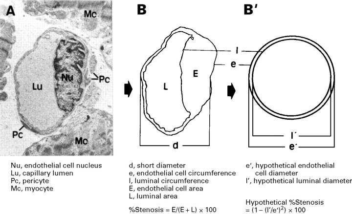
Figure 1 .
Method for ultrastructural morphometry of coronary capillaries. On the electron micrograph of a capillary (A), the circumference of the capillary endothelial cell and of the lumen were traced as shown in panel (B). Panel (B) was then converted to panel (B'), a hypothetical state, using the measured parameters.
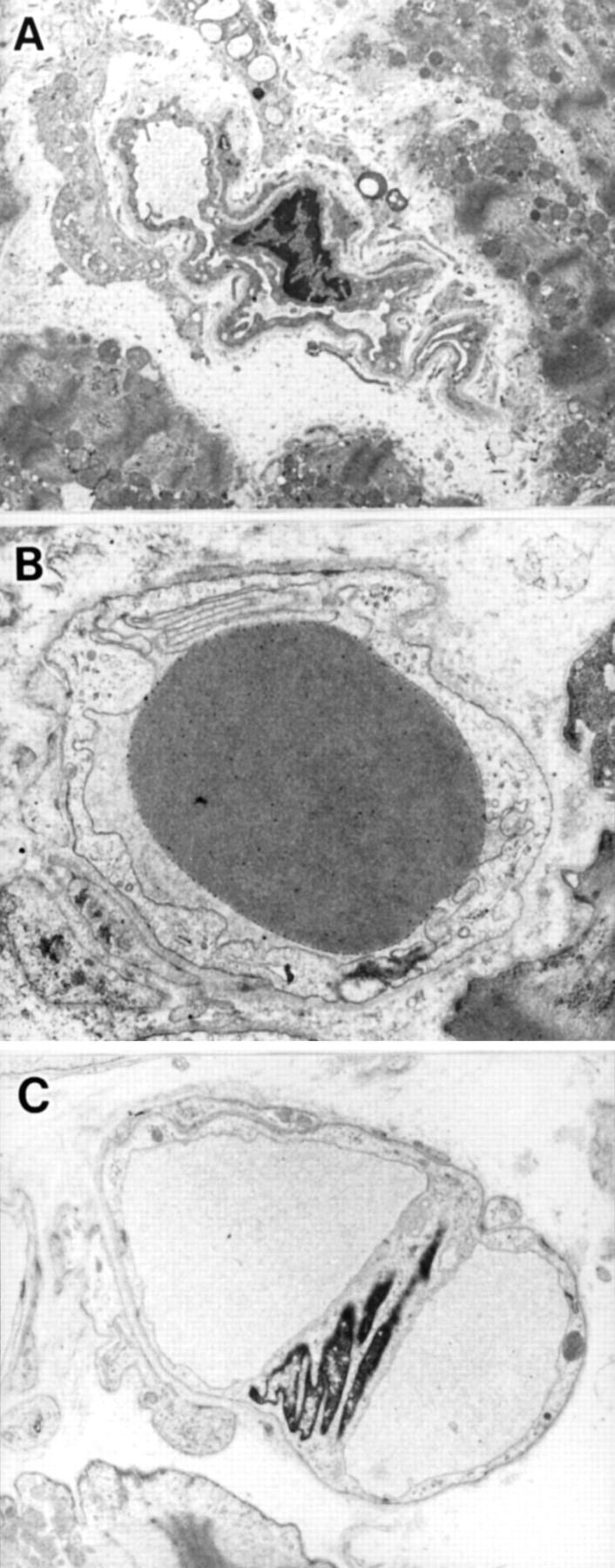
Figure 2 .

(A) Non-round capillaries caused by oblique transection; (B) round but not sectioned at the nucleus level of the capillary endothelial cell; (C) branching capillaries. These were excluded, and only capillaries cut roundly in cross section (the longest and shortest diameter ratio < 2) at the level of the nucleus were examined. (Original magnifications, (A) ×1700; (B) ×5000; (C) ×2500.)
Figure 3 .

(A) Number of the capillaries with abnormal stenosis (> 90 %) in each group. (B) Mean percentage stenosis of the capillaries in each group.
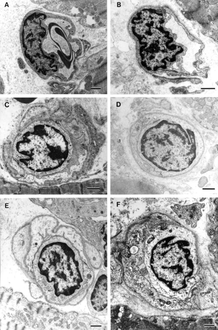
Figure 4 .

Electronmicrographs of the coronary capillaries with various degrees of stenosis. (A) Capillary from a control subject with stenosis of 60%. A profoundly deformed red blood cell is observed in the lumen (original magnification, ×3900). (B) A capillary from a patient with HCM with stenosis of 71% (×5800). (C-F) Severely stenosed capillaries from patients with DCM-like HCM. Stenoses of the capillary lumens are (C) 87%, (D) 98%, (E) 99%, (F) 97%. In (C) and (E), the capillary endothelial cells are unevenly thickened, and almost diffusely in (F). There is no conspicuous thickening of the endothelial cells in (D), but the lumen is slit-like. (Original magnifications: (C and D) ×5000; (E) ×4300; (F) ×4000; bar = 1 µm.)
Selected References
These references are in PubMed. This may not be the complete list of references from this article.
- Aretz H. T., Billingham M. E., Edwards W. D., Factor S. M., Fallon J. T., Fenoglio J. J., Jr, Olsen E. G., Schoen F. J. Myocarditis. A histopathologic definition and classification. Am J Cardiovasc Pathol. 1987 Jan;1(1):3–14. [PubMed] [Google Scholar]
- Baandrup U., Florio R. A., Roters F., Olsen E. G. Electron microscopic investigation of endomyocardial biopsy samples in hypertrophy and cardiomyopathy. A semiquantitative study in 48 patients. Circulation. 1981 Jun;63(6):1289–1298. doi: 10.1161/01.cir.63.6.1289. [DOI] [PubMed] [Google Scholar]
- Edwards W. D. Myocarditis and endomyocardial biopsy. Cardiol Clin. 1984 Nov;2(4):647–656. [PubMed] [Google Scholar]
- Fujiwara H., Kawai C., Hamashima Y. Myocardial fascicle and fiber disarray in 25 mu-thick sections. Circulation. 1979 Jun;59(6):1293–1298. doi: 10.1161/01.cir.59.6.1293. [DOI] [PubMed] [Google Scholar]
- Fujiwara H., Onodera T., Tanaka M., Shirane H., Kato H., Yoshikawa J., Osakada G., Sasayama S., Kawai C. Progression from hypertrophic obstructive cardiomyopathy to typical dilated cardiomyopathy-like features in the end stage. Jpn Circ J. 1984 Nov;48(11):1210–1214. doi: 10.1253/jcj.48.1210. [DOI] [PubMed] [Google Scholar]
- James T. N., Marshall T. K. De subitaneis mortibus. XII. Asymmetrical hypertrophy of the heart. Circulation. 1975 Jun;51(6):1149–1166. doi: 10.1161/01.cir.51.6.1149. [DOI] [PubMed] [Google Scholar]
- James T. N. Small arteries of the heart. Circulation. 1977 Jul;56(1):2–14. doi: 10.1161/01.cir.56.1.2. [DOI] [PubMed] [Google Scholar]
- Kawamura K., James T. N. Comparative ultrastructure of cellular junctions in working myocardium and the conduction system under normal and pathologic conditions. J Mol Cell Cardiol. 1971 Sep;3(1):31–60. doi: 10.1016/0022-2828(71)90031-9. [DOI] [PubMed] [Google Scholar]
- Kloner R. A., Rude R. E., Carlson N., Maroko P. R., DeBoer L. W., Braunwald E. Ultrastructural evidence of microvascular damage and myocardial cell injury after coronary artery occlusion: which comes first? Circulation. 1980 Nov;62(5):945–952. doi: 10.1161/01.cir.62.5.945. [DOI] [PubMed] [Google Scholar]
- Maron B. J., Epstein S. E., Roberts W. C. Hypertrophic cardiomyopathy and transmural myocardial infarction without significant atherosclerosis of the extramural coronary arteries. Am J Cardiol. 1979 Jun;43(6):1086–1102. doi: 10.1016/0002-9149(79)90139-5. [DOI] [PubMed] [Google Scholar]
- Maron B. J., Ferrans V. J., Roberts W. C. Ultrastructural features of degenerated cardiac muscle cells in patients with cardiac hypertrophy. Am J Pathol. 1975 Jun;79(3):387–434. [PMC free article] [PubMed] [Google Scholar]
- Maron B. J., Wolfson J. K., Epstein S. E., Roberts W. C. Intramural ("small vessel") coronary artery disease in hypertrophic cardiomyopathy. J Am Coll Cardiol. 1986 Sep;8(3):545–557. doi: 10.1016/s0735-1097(86)80181-4. [DOI] [PubMed] [Google Scholar]
- Mosseri M., Schaper J., Admon D., Hasin Y., Gotsman M. S., Sapoznikov D., Pickering J. G., Yarom R. Coronary capillaries in patients with congestive cardiomyopathy or angina pectoris with patent main coronary arteries. Ultrastructural morphometry of endomyocardial biopsy samples. Circulation. 1991 Jul;84(1):203–210. doi: 10.1161/01.cir.84.1.203. [DOI] [PubMed] [Google Scholar]
- Olmesdahl P. J., Gregory M. A., Cameron E. W. Ultrastructural artefacts in biopsied normal myocardium and their relevance to myocardial biopsy in man. Thorax. 1979 Feb;34(1):82–90. doi: 10.1136/thx.34.1.82. [DOI] [PMC free article] [PubMed] [Google Scholar]
- Onodera T., Fujiwara H., Tanaka M., Wu D. J., Hamashima Y., Kawai C. Familial hypertrophic cardiomyopathy mimicing typical dilated cardiomyopathy. Jpn Circ J. 1986 Jul;50(7):614–618. doi: 10.1253/jcj.50.614. [DOI] [PubMed] [Google Scholar]
- Pichard A. D., Meller J., Teichholz L. E., Lipnik S., Gorlin R., Herman M. V. Septal perforator compression (narrowing) in idiopathic hypertrophic subaortic stenosis. Am J Cardiol. 1977 Sep;40(3):310–314. doi: 10.1016/0002-9149(77)90151-5. [DOI] [PubMed] [Google Scholar]
- Rakusan K. Quantitative morphology of capillaries of the heart. Number of capillaries in animal and human hearts under normal and pathological conditions. Methods Achiev Exp Pathol. 1971;5:272–286. [PubMed] [Google Scholar]
- Sonnenblick E. H. Correlation of myocardial ultrastructure and function. Circulation. 1968 Jul;38(1):29–44. doi: 10.1161/01.cir.38.1.29. [DOI] [PubMed] [Google Scholar]
- Tanaka M., Fujiwara H., Onodera T., Wu D. J., Matsuda M., Hamashima Y., Kawai C. Quantitative analysis of narrowings of intramyocardial small arteries in normal hearts, hypertensive hearts, and hearts with hypertrophic cardiomyopathy. Circulation. 1987 Jun;75(6):1130–1139. doi: 10.1161/01.cir.75.6.1130. [DOI] [PubMed] [Google Scholar]
- Warnes C. A., Maron B. J., Roberts W. C. Massive cardiac ventricular scarring in first-degree relatives with hypertrophic cardiomyopathy. Am J Cardiol. 1984 Dec 1;54(10):1377–1379. doi: 10.1016/s0002-9149(84)80108-3. [DOI] [PubMed] [Google Scholar]
- Weiss M. B., Ellis K., Sciacca R. R., Johnson L. L., Schmidt D. H., Cannon P. J. Myocardial blood flow in congestive and hypertrophic cardiomyopathy: relationship to peak wall stress and mean velocity of circumferential fiber shortening. Circulation. 1976 Sep;54(3):484–494. doi: 10.1161/01.cir.54.3.484. [DOI] [PubMed] [Google Scholar]
- Yutani C., Imakita M., Ishibashi-Ueda H., Hatanaka K., Nagata S., Sakakibara H., Nimura Y. Three autopsy cases of progression to left ventricular dilatation in patients with hypertrophic cardiomyopathy. Am Heart J. 1985 Mar;109(3 Pt 1):545–553. doi: 10.1016/0002-8703(85)90561-7. [DOI] [PubMed] [Google Scholar]


